Best Drawing Technique When Using Microscope ?
The best drawing technique when using a microscope is to start by adjusting the focus and lighting to obtain a clear and well-illuminated image of the specimen. Then, carefully observe the details of the specimen under the microscope and mentally visualize the desired drawing. It is important to use a fine-tipped pencil or a technical pen to accurately capture the intricate details. Begin by lightly sketching the outlines and major features of the specimen, gradually adding more details as you progress. Pay attention to proportions, textures, and shading to create a realistic representation. It is recommended to work slowly and patiently, making small adjustments and corrections as needed. Additionally, using a grid or ruler can help maintain accuracy and scale in the drawing. Finally, remember to label important structures or parts of the specimen to provide clarity and context.
1、 Microscopic observation and sketching techniques for accurate representation.
The best drawing technique when using a microscope is to employ microscopic observation and sketching techniques for accurate representation. This approach ensures that the details and features observed under the microscope are accurately depicted in the drawing.
To begin, it is crucial to have a clear understanding of the specimen being observed. Take the time to carefully observe and study the specimen under the microscope, noting its shape, color, texture, and any other relevant characteristics. This will help in accurately representing these features in the drawing.
When sketching, it is important to use a fine-tipped pencil or pen to capture the intricate details of the specimen. Start by outlining the basic shape of the specimen and then gradually add more details, paying close attention to the proportions and relationships between different parts. It is advisable to work slowly and patiently, making sure to accurately represent the observed features.
Additionally, it is beneficial to use shading techniques to create depth and dimension in the drawing. This can be achieved by carefully observing the lighting and shadows present in the specimen and replicating them in the drawing. Shading can help in accurately representing the three-dimensional nature of the specimen, making the drawing more realistic.
Furthermore, it is important to constantly refer back to the microscope while sketching to ensure accuracy. This will help in capturing any minute details that may be missed otherwise.
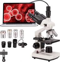
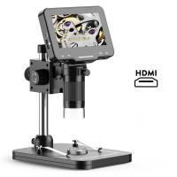
In recent years, advancements in technology have provided new tools for microscopic observation and sketching. Digital microscopes and imaging software allow for more precise and detailed observations, which can be directly translated into digital drawings. This can be particularly useful for scientific research and documentation.
In conclusion, the best drawing technique when using a microscope is to employ microscopic observation and sketching techniques for accurate representation. By carefully observing the specimen, using fine-tipped tools, incorporating shading techniques, and constantly referring back to the microscope, one can create a drawing that faithfully represents the observed features. The latest advancements in technology have also provided new opportunities for digital sketching, further enhancing the accuracy and precision of microscopic drawings.
2、 Staining methods to enhance visibility and contrast in microscopic drawings.
The best drawing technique when using a microscope is to utilize staining methods to enhance visibility and contrast in microscopic drawings. Staining is a technique that involves applying specific dyes or chemicals to a specimen, which helps to highlight certain structures or components of the sample. This method allows for better visualization and differentiation of various cellular or tissue components, making it easier to accurately depict them in a drawing.
There are several staining methods available, each with its own advantages and applications. For example, hematoxylin and eosin (H&E) staining is commonly used in histology to differentiate between different types of cells and tissues. This staining technique imparts a blue color to the nuclei of cells and a pink color to the cytoplasm, allowing for clear distinction between different cellular structures.
Other staining methods, such as immunohistochemistry, can be used to specifically label certain proteins or molecules within a sample. This technique involves using antibodies that are tagged with a fluorescent or enzymatic marker, which can then be visualized under a microscope. Immunohistochemistry staining is particularly useful in identifying specific cell types or markers within a tissue sample.
In recent years, advancements in staining techniques have been made, such as the development of multiplex staining methods. These methods allow for the simultaneous visualization of multiple targets within a single sample, providing a more comprehensive understanding of cellular interactions and structures.
In conclusion, staining methods are the best drawing technique when using a microscope as they enhance visibility and contrast in microscopic drawings. These techniques allow for accurate depiction of cellular structures and components, aiding in the understanding and communication of microscopic observations. The latest advancements in staining methods, such as multiplex staining, further enhance the capabilities of microscopic drawings by enabling the visualization of multiple targets within a single sample.

3、 Proper lighting techniques for microscope-based drawing.
The best drawing technique when using a microscope involves a combination of proper lighting techniques and attention to detail. Proper lighting is crucial for accurately capturing the specimen's features and ensuring that the drawing is clear and well-defined.
One of the most important lighting techniques is to adjust the microscope's light source to achieve optimal illumination. This can be done by adjusting the intensity of the light or using filters to enhance contrast. It is essential to avoid excessive brightness, as it can wash out details, and insufficient lighting, as it can make the specimen appear dull and indistinct.
Another important aspect is the angle of illumination. By adjusting the angle of the light source, different features of the specimen can be highlighted, allowing for a more comprehensive and accurate drawing. Experimenting with different angles can reveal hidden details and provide a more comprehensive understanding of the specimen's structure.
Additionally, using a condenser can help focus the light onto the specimen, improving the clarity and visibility of the details. Adjusting the condenser's aperture can control the amount of light that reaches the specimen, allowing for better contrast and definition.
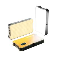
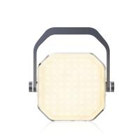
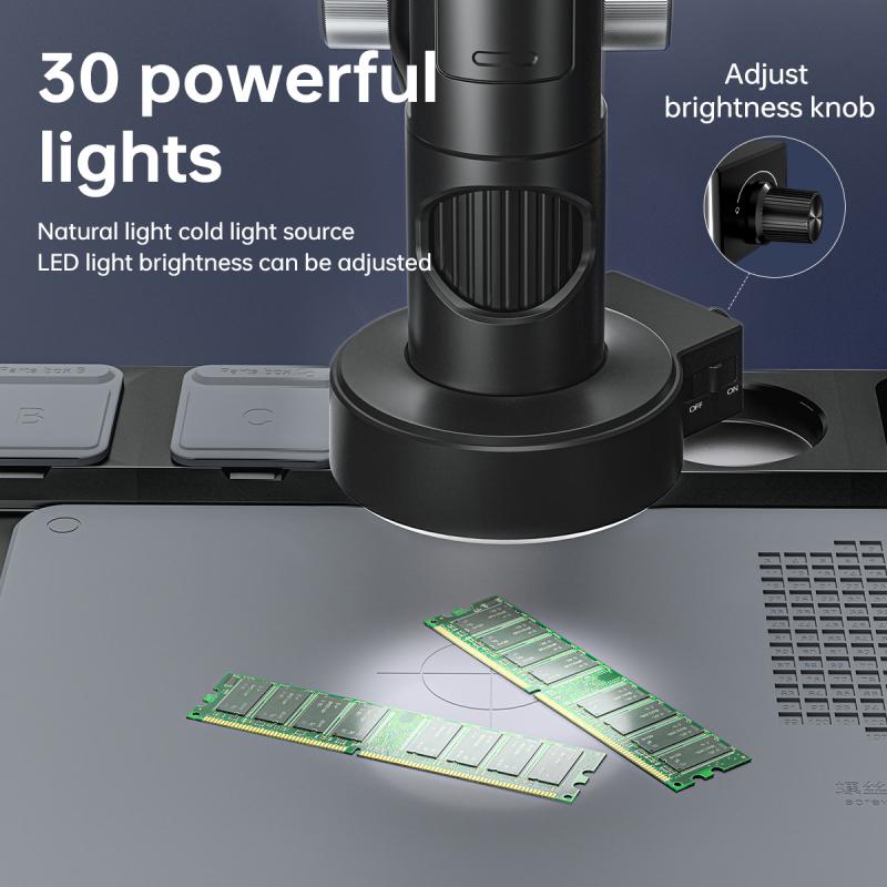
In recent years, advancements in microscope technology have introduced new lighting techniques, such as LED illumination. LED lights offer several advantages, including longer lifespan, lower energy consumption, and adjustable color temperature. These advancements have further improved the quality of microscope-based drawings by providing more control over lighting conditions.
In conclusion, the best drawing technique when using a microscope involves proper lighting techniques. By adjusting the light source's intensity, angle, and using tools like condensers, one can achieve optimal illumination and capture the specimen's features accurately. The latest advancements in LED lighting have further enhanced the quality of microscope-based drawings, providing more control and flexibility in lighting conditions.

4、 Tips for capturing fine details and textures in microscope drawings.
The best drawing technique when using a microscope involves a combination of observation skills, attention to detail, and artistic techniques. Here are some tips for capturing fine details and textures in microscope drawings:
1. Observe carefully: Take your time to observe the specimen under the microscope. Pay attention to the intricate details, textures, and patterns. Use the microscope's focus and magnification adjustments to explore different areas of interest.
2. Use proper lighting: Adjust the lighting conditions to enhance the visibility of the specimen. Experiment with different lighting angles and intensities to bring out the fine details and textures.
3. Start with a rough sketch: Begin by making a rough outline of the specimen on your drawing paper. This will help you establish the overall shape and proportions before adding the finer details.
4. Use a variety of pencils: Experiment with different pencil grades to achieve varying levels of shading and texture. Use lighter pencils (e.g., 2H or HB) for lighter areas and darker pencils (e.g., 2B or 4B) for darker areas. This will help you create depth and dimension in your drawing.
5. Employ shading techniques: Use techniques like hatching, cross-hatching, stippling, and blending to capture the textures and shading of the specimen. These techniques can help you recreate the intricate patterns and details observed under the microscope.
6. Pay attention to composition: Consider the composition of your drawing. Arrange the specimen in a visually appealing way and use negative space effectively to highlight the subject.
7. Practice regularly: Like any skill, drawing under a microscope requires practice. Regularly sketching different specimens will improve your observation skills and help you develop your own unique style.
In the latest point of view, advancements in digital microscopy have opened up new possibilities for capturing fine details and textures. Digital microscopes allow for real-time imaging and can be connected to a computer or tablet, enabling artists to directly draw on a digital canvas. This technology offers advantages such as the ability to zoom in and out, adjust lighting and contrast, and easily undo or modify drawings. Artists can also take advantage of various digital tools and software to enhance their microscope drawings further. However, traditional drawing techniques remain valuable and can be combined with digital methods to create stunning microscope drawings.
