Can Bacteria Be Seen With A Light Microscope ?
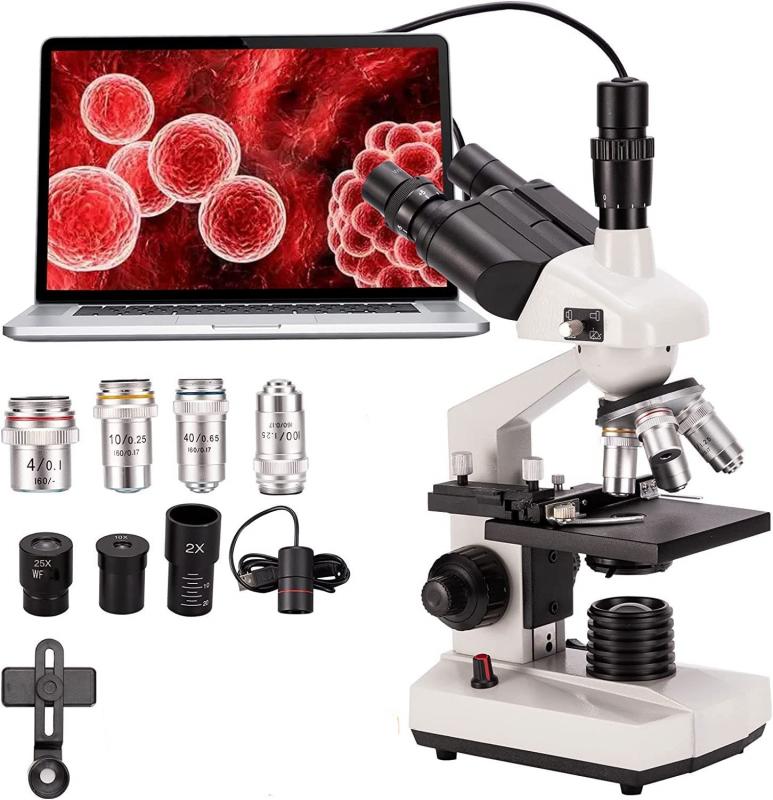
Yes, bacteria can be seen with a light microscope. Light microscopes use visible light to illuminate the specimen and magnify it for observation. Bacteria are typically small in size, ranging from 0.2 to 10 micrometers in diameter, which makes them suitable for observation under a light microscope. However, it is important to note that some bacteria may require staining techniques to enhance their visibility and contrast under the microscope.
1、 Bacterial morphology and size under light microscopy
Yes, bacteria can be seen with a light microscope. Light microscopy is a widely used technique for studying bacterial morphology and size. Bacteria are microscopic organisms, and their size typically ranges from 0.2 to 10 micrometers. This makes them well-suited for observation under a light microscope, which has a resolution limit of around 0.2 micrometers.
When using a light microscope, bacteria can be visualized by staining them with dyes that enhance contrast and make them more visible. Commonly used stains include crystal violet, methylene blue, and Gram stain. These stains bind to different components of the bacterial cell, such as the cell wall or DNA, allowing for better visualization and differentiation of bacterial species.
However, it is important to note that the resolution of a light microscope is limited by the wavelength of visible light. This means that some smaller bacteria may not be clearly visible under a light microscope, and their detailed structures may not be discernible. In such cases, higher-resolution techniques like electron microscopy may be necessary.
It is worth mentioning that advancements in microscopy techniques have led to the development of super-resolution microscopy, which can overcome the diffraction limit of light and provide higher resolution images. This has allowed researchers to study bacterial structures and processes in more detail. However, super-resolution microscopy is still a relatively new and specialized technique, and its widespread use in bacterial research is limited.
In conclusion, while light microscopy is a valuable tool for studying bacterial morphology and size, its resolution is limited, and some smaller bacteria may not be clearly visible. Researchers may need to employ other techniques, such as electron microscopy or super-resolution microscopy, to obtain more detailed information about bacterial structures.
2、 Staining techniques for visualizing bacteria with a light microscope
Yes, bacteria can be seen with a light microscope. However, due to their small size, they are not easily visible without the use of staining techniques. Staining techniques enhance the contrast between the bacteria and their surroundings, making them easier to observe under a light microscope.
There are several staining techniques commonly used to visualize bacteria. One of the most widely used techniques is the Gram staining method, developed by Hans Christian Gram in 1884. This technique involves staining bacteria with crystal violet, followed by iodine treatment, decolorization with alcohol or acetone, and counterstaining with safranin. Gram-positive bacteria retain the crystal violet stain and appear purple, while Gram-negative bacteria lose the stain and appear pink.
Another commonly used staining technique is the acid-fast staining method, which is used to identify bacteria of the genus Mycobacterium, including the causative agent of tuberculosis. Acid-fast staining involves staining bacteria with carbol fuchsin, followed by acid-alcohol decolorization and counterstaining with methylene blue. Acid-fast bacteria retain the carbol fuchsin stain and appear red, while other bacteria appear blue.
Fluorescent staining techniques have also been developed, which involve using fluorescent dyes that bind specifically to certain components of bacterial cells. These dyes emit fluorescence when exposed to specific wavelengths of light, allowing for the visualization of bacteria under a fluorescent microscope.
It is important to note that while staining techniques greatly enhance the visibility of bacteria, they can also introduce artifacts and alter the natural characteristics of the cells. Therefore, it is crucial to use staining techniques judiciously and interpret the results with caution.
In recent years, advancements in microscopy technology, such as confocal microscopy and super-resolution microscopy, have further improved the visualization of bacteria. These techniques allow for higher resolution and three-dimensional imaging of bacterial cells, providing researchers with more detailed information about their structure and behavior.
In conclusion, while bacteria can be seen with a light microscope, staining techniques are necessary to enhance their visibility. These techniques, such as Gram staining and acid-fast staining, have been widely used for many years. Additionally, newer microscopy technologies have further improved our ability to visualize bacteria and study their intricate details.
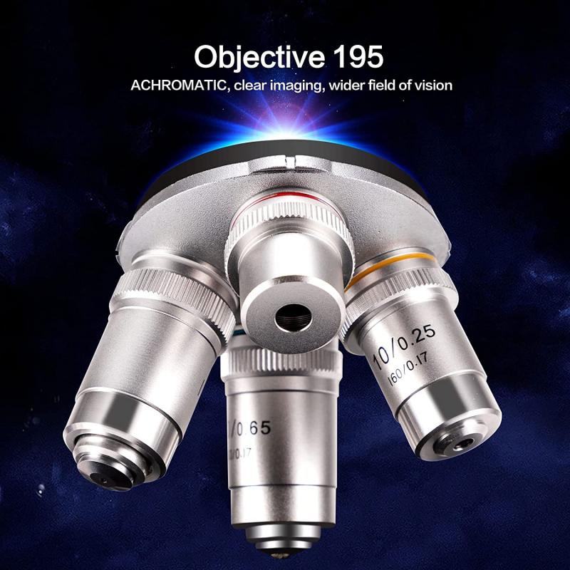
3、 Limitations of light microscopy in observing bacteria
Yes, bacteria can be seen with a light microscope. Light microscopy is a widely used technique for observing bacteria due to its simplicity, affordability, and accessibility. By using a light microscope, scientists can visualize bacteria and study their morphology, size, arrangement, and motility.
However, there are limitations to using light microscopy in observing bacteria. One major limitation is the resolution of the microscope. The resolution of a light microscope is limited by the wavelength of visible light, which is around 400-700 nanometers. This means that bacteria smaller than this limit may not be clearly visible under a light microscope. Some bacteria, such as certain species of Mycoplasma, are extremely small and may require more advanced techniques like electron microscopy for visualization.
Another limitation is the lack of contrast in bacterial samples. Bacteria are often transparent and lack pigmentation, making them difficult to distinguish from their surroundings. To overcome this, scientists use staining techniques to enhance contrast and make bacteria more visible under a light microscope. Stains like Gram stain and acid-fast stain can help differentiate bacteria based on their cell wall composition.
Furthermore, light microscopy may not provide enough detail to identify specific bacterial species. While it can reveal general characteristics, such as shape and arrangement, it may not be sufficient for species-level identification. Molecular techniques, such as DNA sequencing, are often required for accurate identification.
In recent years, advancements in light microscopy techniques have improved the visualization of bacteria. Techniques like phase-contrast microscopy, dark-field microscopy, and fluorescence microscopy have enhanced contrast and allowed for better visualization of bacteria. Additionally, the development of super-resolution microscopy techniques has pushed the limits of resolution, enabling the visualization of smaller bacterial structures.
In conclusion, while light microscopy can be used to observe bacteria, it has limitations in terms of resolution, contrast, and species-level identification. However, with the continuous advancements in microscopy techniques, researchers are able to overcome some of these limitations and gain a better understanding of the microbial world.
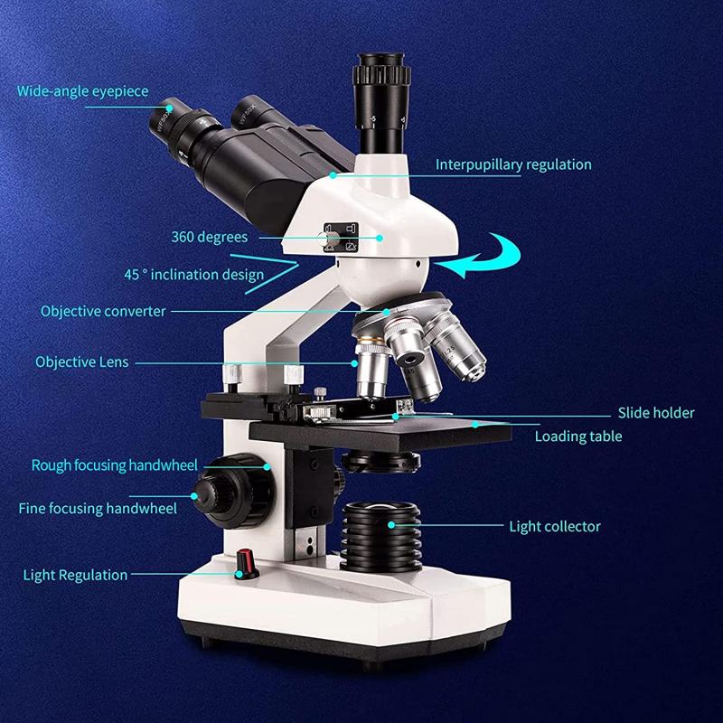
4、 Advances in microscopy for improved visualization of bacteria
Yes, bacteria can be seen with a light microscope. Light microscopy has been a fundamental tool in microbiology for over a century, allowing scientists to observe and study bacteria. However, it is important to note that bacteria are extremely small, typically ranging in size from 0.2 to 10 micrometers, which is at the limit of resolution for a light microscope.
To visualize bacteria using a light microscope, scientists often employ staining techniques. Stains such as crystal violet, methylene blue, or Gram stain can be used to enhance the contrast between the bacteria and their surroundings, making them easier to see. These stains can also provide information about the bacterial cell structure and composition.
Advances in microscopy technology have greatly improved the visualization of bacteria. For instance, the development of fluorescence microscopy has allowed scientists to label specific components of bacterial cells with fluorescent dyes or proteins, enabling the visualization of specific structures or molecules within the bacteria. This technique has revolutionized our understanding of bacterial cell biology and has been instrumental in studying processes such as bacterial division, protein localization, and gene expression.
Furthermore, the advent of confocal microscopy has provided three-dimensional imaging capabilities, allowing researchers to visualize bacteria in complex environments such as biofilms or host tissues. Confocal microscopy uses a laser to scan through the sample, collecting images at different depths and reconstructing them into a three-dimensional image.
In recent years, super-resolution microscopy techniques, such as stimulated emission depletion (STED) microscopy and structured illumination microscopy (SIM), have pushed the limits of resolution beyond the diffraction limit of light. These techniques have allowed scientists to visualize bacterial structures and processes at the nanoscale level, providing unprecedented insights into the intricate details of bacterial biology.
In conclusion, while bacteria can be seen with a light microscope, advances in microscopy techniques have significantly improved our ability to visualize and study bacteria. These advancements have expanded our understanding of bacterial cell biology and have the potential to contribute to the development of new strategies for combating bacterial infections.
