Can Mitochondria Be Seen Under A Light Microscope ?
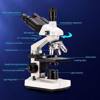
Yes, mitochondria can be seen under a light microscope. However, they are quite small and may not be easily visible without staining or using specialized techniques to enhance their visibility.
1、 Mitochondria: Structure and Function
Yes, mitochondria can be seen under a light microscope. However, due to their small size and transparent nature, they are not easily visible without staining or using specialized techniques.
Mitochondria are double-membraned organelles found in most eukaryotic cells. They are responsible for producing energy in the form of adenosine triphosphate (ATP) through a process called cellular respiration. The inner membrane of mitochondria contains proteins and enzymes that are involved in this energy production.
Under a light microscope, mitochondria appear as small, oval-shaped structures. They can be stained with dyes such as MitoTracker or Rhodamine 123, which specifically target the mitochondria and make them more visible. These stains bind to the mitochondria and emit fluorescence when excited by light, allowing researchers to observe and study their structure and function.
In recent years, advancements in microscopy techniques have allowed for better visualization of mitochondria. Super-resolution microscopy, such as stimulated emission depletion (STED) microscopy and structured illumination microscopy (SIM), can provide higher resolution images of mitochondria, revealing more details about their structure and organization within the cell.
It is important to note that while light microscopy can provide valuable information about the size, shape, and distribution of mitochondria, it has limitations in terms of resolution. To study the ultrastructure and finer details of mitochondria, electron microscopy is often used. Electron microscopy allows for higher magnification and resolution, enabling researchers to observe the internal structure of mitochondria, including the cristae and matrix.
In conclusion, while mitochondria can be observed under a light microscope, staining techniques and advanced microscopy methods are often required to enhance their visibility and study their structure and function in more detail.

2、 Mitochondrial Dynamics and Fission-Fusion
Yes, mitochondria can be seen under a light microscope. However, due to their small size and transparent nature, they are not easily visible without specific staining techniques or fluorescent labeling. Mitochondria are typically stained with dyes such as MitoTracker or Rhodamine 123, which specifically target the mitochondria and make them more visible under a light microscope.
Mitochondrial dynamics and fission-fusion refer to the continuous process of mitochondrial division (fission) and fusion, which play crucial roles in maintaining mitochondrial function and cellular homeostasis. These processes are essential for regulating mitochondrial size, shape, distribution, and quality control.
Recent advancements in microscopy techniques, such as super-resolution microscopy, have provided a more detailed understanding of mitochondrial dynamics. These techniques allow researchers to visualize and study the intricate processes of mitochondrial fission and fusion at a higher resolution. Super-resolution microscopy has revealed that mitochondria undergo dynamic changes in shape and size, forming interconnected networks within cells.
Additionally, live-cell imaging techniques have enabled the observation of mitochondrial dynamics in real-time. This has provided insights into the mechanisms underlying mitochondrial fission and fusion, as well as their functional implications in cellular processes such as energy production, apoptosis, and cellular signaling.
In conclusion, while mitochondria can be observed under a light microscope with the aid of specific staining techniques, recent advancements in microscopy have allowed for a more detailed and dynamic understanding of mitochondrial dynamics and fission-fusion. These techniques have provided valuable insights into the role of mitochondria in cellular function and have opened up new avenues for further research in this field.

3、 Mitochondrial DNA and Replication
Yes, mitochondria can be seen under a light microscope. However, due to their small size and transparent nature, they are not easily visible without staining or using specialized techniques. Mitochondria are double-membraned organelles found in most eukaryotic cells, responsible for producing energy in the form of ATP through cellular respiration. They contain their own DNA, known as mitochondrial DNA (mtDNA), which is separate from the nuclear DNA.
To visualize mitochondria under a light microscope, researchers often use dyes or stains that specifically target these organelles. One commonly used stain is MitoTracker, which selectively accumulates in mitochondria and emits a fluorescent signal when excited by light. This allows researchers to observe and study the morphology, distribution, and dynamics of mitochondria in living cells.
In recent years, advancements in microscopy techniques have further improved the visualization of mitochondria. Super-resolution microscopy, such as stimulated emission depletion (STED) microscopy and structured illumination microscopy (SIM), can achieve resolutions beyond the diffraction limit of light. These techniques enable researchers to observe mitochondria with higher clarity and detail, providing insights into their structure and function.
It is worth noting that the ability to visualize mitochondria under a light microscope is limited by the resolution of the microscope and the techniques used. Electron microscopy, which uses a beam of electrons instead of light, provides even higher resolution and can reveal more intricate details of mitochondria. However, it requires more complex sample preparation and is not as readily accessible as light microscopy.
In conclusion, while mitochondria can be seen under a light microscope, their visualization often requires staining or specialized techniques. Advancements in microscopy have enhanced our understanding of mitochondria, but electron microscopy remains the gold standard for studying their ultrastructure.

4、 Mitochondrial Biogenesis and Protein Import
Yes, mitochondria can be seen under a light microscope. However, due to their small size and transparent nature, they are not easily visible without staining or specific techniques. Mitochondria are typically stained with dyes such as MitoTracker or Rhodamine 123, which specifically target the mitochondria and make them more visible under a light microscope.
Mitochondrial biogenesis, the process by which new mitochondria are formed, is a complex and highly regulated process. It involves the replication of mitochondrial DNA, the synthesis of mitochondrial proteins, and their import into the mitochondria. The import of proteins into mitochondria is a crucial step in maintaining mitochondrial function and is tightly regulated.
Recent studies have shed light on the mechanisms involved in mitochondrial protein import. It has been found that proteins destined for the mitochondria are synthesized in the cytosol and then targeted to the mitochondria by specific targeting sequences. These targeting sequences are recognized by receptors on the mitochondrial outer membrane, which facilitate the import of the proteins into the mitochondria.
Furthermore, it has been discovered that the import of proteins into mitochondria is not a one-step process but involves multiple steps and protein complexes. These complexes, such as the translocase of the outer membrane (TOM) and the translocase of the inner membrane (TIM), work together to ensure the efficient import of proteins into the mitochondria.
In conclusion, while mitochondria can be seen under a light microscope with the use of specific staining techniques, the study of mitochondrial biogenesis and protein import requires more advanced techniques such as electron microscopy and molecular biology approaches. The latest research has provided valuable insights into the mechanisms involved in mitochondrial protein import, highlighting the complexity and importance of this process in maintaining mitochondrial function.


