Can They See Coesliac Disease On Endoscope ?
Yes, coeliac disease can be seen on an endoscope.
1、 Endoscopic findings in celiac disease: mucosal abnormalities and atrophy.
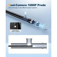
Yes, endoscopy is an important diagnostic tool for detecting and evaluating the presence of celiac disease. During an endoscopy, a flexible tube with a camera at the end, called an endoscope, is inserted through the mouth and into the small intestine to examine the mucosal lining.
Endoscopic findings in celiac disease typically include mucosal abnormalities and atrophy. The mucosal lining of the small intestine may appear flattened, with a loss of the normal finger-like projections called villi. This is known as villous atrophy and is a characteristic feature of celiac disease. The severity of villous atrophy can vary, and it is often graded on a scale from Marsh 0 (normal) to Marsh 3 (severe atrophy).
In addition to villous atrophy, other mucosal abnormalities may be observed during endoscopy. These can include increased lymphocytes (lymphocytic infiltration) in the epithelium, crypt hyperplasia (increased cell growth in the intestinal crypts), and an increase in intraepithelial lymphocytes.
It is important to note that while endoscopy can provide valuable information, it is not the only diagnostic tool for celiac disease. A definitive diagnosis requires a combination of clinical symptoms, blood tests (such as serology for specific antibodies), and histological examination of small intestinal biopsies obtained during endoscopy.
The latest point of view on endoscopic findings in celiac disease suggests that endoscopy with duodenal biopsies is still considered the gold standard for diagnosis. However, there is ongoing research to explore alternative diagnostic methods, such as non-invasive tests that can detect celiac disease without the need for endoscopy. These tests include serological markers and genetic testing. While these non-invasive tests show promise, they are not yet widely used in clinical practice and further validation is needed.
In conclusion, endoscopy plays a crucial role in the diagnosis of celiac disease by allowing direct visualization of mucosal abnormalities and atrophy in the small intestine. It provides important information that, when combined with other diagnostic tools, helps confirm the presence of celiac disease and guide appropriate management strategies.
2、 Marsh classification: grading the severity of celiac disease histologically.
Yes, coeliac disease can be seen on an endoscope through histological examination. The Marsh classification system is commonly used to grade the severity of celiac disease based on histological findings. This classification system helps in diagnosing and monitoring the disease.
The Marsh classification system consists of several stages, ranging from Marsh 0 to Marsh 4, each representing different levels of damage to the small intestine. In Marsh 0, there is no visible damage to the intestinal lining, but there may be an increased number of immune cells present. As the severity progresses, Marsh 1 shows an increase in intraepithelial lymphocytes, Marsh 2 shows the presence of crypt hyperplasia, Marsh 3 shows villous atrophy, and Marsh 4 represents complete villous atrophy.
During an endoscopy, a small tissue sample (biopsy) is taken from the small intestine and examined under a microscope to determine the Marsh classification. The biopsy helps to identify the characteristic changes in the intestinal lining associated with celiac disease, such as inflammation, damage to the villi, and increased immune cell infiltration.
It is important to note that the Marsh classification system has been subject to some debate and modifications over the years. The latest point of view suggests that the Marsh classification may not fully capture the heterogeneity of celiac disease and its clinical implications. Some experts argue that additional factors, such as the presence of specific antibodies and genetic markers, should also be considered for a comprehensive diagnosis and management of the disease.
In conclusion, endoscopic examination with histological analysis can detect and classify the severity of celiac disease using the Marsh classification system. However, it is essential to consider the latest perspectives and additional diagnostic markers for a more comprehensive understanding of the disease.
3、 Endoscopic markers of gluten exposure in celiac disease patients.
Yes, endoscopy can be used to detect and evaluate the presence of celiac disease in patients. Celiac disease is an autoimmune disorder triggered by the ingestion of gluten, a protein found in wheat, barley, and rye. The disease causes damage to the small intestine, leading to various gastrointestinal symptoms and nutrient malabsorption.
During an endoscopy, a thin, flexible tube with a camera at the end (endoscope) is inserted through the mouth and into the small intestine. This allows the doctor to visually examine the lining of the intestine and take biopsies if necessary. In the case of celiac disease, the endoscopy can reveal characteristic markers of gluten exposure.
The most notable marker seen during endoscopy is the presence of villous atrophy, which refers to the flattening of the finger-like projections (villi) in the small intestine. Villous atrophy is a hallmark of celiac disease and indicates significant damage to the intestinal lining. Other markers that may be observed include inflammation, ulceration, and a mosaic pattern on the mucosal surface.
It is important to note that endoscopy alone cannot definitively diagnose celiac disease. A confirmed diagnosis requires a combination of clinical symptoms, blood tests (such as serology for specific antibodies), and histological examination of intestinal biopsies obtained during endoscopy. Additionally, it is recommended that patients continue consuming gluten-containing foods prior to the procedure to ensure accurate results.
The latest point of view on endoscopic markers of gluten exposure in celiac disease patients emphasizes the importance of a thorough evaluation of the small intestine during endoscopy. This includes careful examination of the duodenal bulb, as well as multiple biopsies from different areas of the small intestine to increase diagnostic accuracy. Furthermore, advancements in endoscopic imaging techniques, such as high-definition endoscopy and confocal laser endomicroscopy, have shown promise in enhancing the detection and characterization of celiac disease-related changes in the intestinal mucosa.
In conclusion, endoscopy plays a crucial role in the diagnosis and evaluation of celiac disease. While endoscopic markers of gluten exposure, such as villous atrophy, can be observed during the procedure, a comprehensive diagnostic approach combining clinical, serological, and histological findings is necessary for an accurate diagnosis.
4、 Potential complications of celiac disease visible on endoscopy.
Yes, celiac disease can be seen on an endoscope. Endoscopy is a commonly used procedure to diagnose celiac disease and assess its potential complications. During an endoscopy, a thin, flexible tube with a camera on the end (endoscope) is inserted through the mouth and into the small intestine. This allows the doctor to visualize the lining of the small intestine and look for any abnormalities.
In the case of celiac disease, the endoscopy can reveal characteristic findings such as a flattened or scalloped appearance of the small intestine lining, known as villous atrophy. The villi are tiny finger-like projections that line the small intestine and aid in nutrient absorption. In celiac disease, these villi become damaged and flattened due to the immune system's reaction to gluten, a protein found in wheat, barley, and rye.
Additionally, the endoscopy may also help identify potential complications of celiac disease. These complications can include ulcers, strictures (narrowing of the intestine), or malignancies such as lymphoma. However, it is important to note that these complications are relatively rare in celiac disease.
It is worth mentioning that the latest point of view suggests that a biopsy of the small intestine is still considered the gold standard for diagnosing celiac disease, even though endoscopy can provide visual evidence of the disease. The biopsy involves taking small tissue samples from the small intestine during the endoscopy procedure and examining them under a microscope for the presence of villous atrophy or other signs of celiac disease.
In conclusion, endoscopy can indeed reveal the presence of celiac disease and its potential complications. However, a biopsy is still necessary for an accurate diagnosis. If you suspect you may have celiac disease, it is important to consult with a healthcare professional for proper evaluation and diagnosis.











There are no comments for this blog.