Can You See Bacteria With A Microscope ?
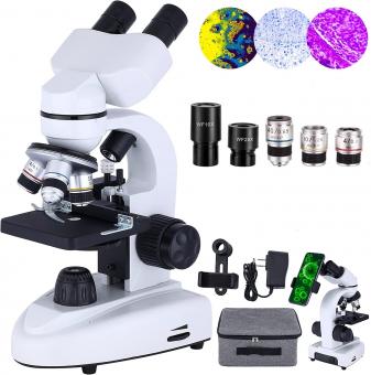
Yes, bacteria can be seen with a microscope. However, due to their small size, they require a high-powered microscope to be visible. Bacteria are typically between 0.5 and 5 micrometers in size, which is much smaller than the human eye can see. Therefore, a microscope is necessary to observe them. There are different types of microscopes that can be used to view bacteria, including light microscopes, electron microscopes, and fluorescence microscopes. The type of microscope used depends on the specific research question and the level of detail required.
1、 Bacterial Size and Shape

Yes, bacteria can be seen with a microscope. In fact, the invention of the microscope in the 17th century allowed scientists to observe and study bacteria for the first time. Bacteria are typically too small to be seen with the naked eye, ranging in size from 0.2 to 10 micrometers in diameter. However, with the use of a light microscope or an electron microscope, bacteria can be visualized and studied in detail.
Bacterial size and shape can vary greatly depending on the species. Some bacteria are spherical in shape, known as cocci, while others are rod-shaped, known as bacilli. There are also spiral-shaped bacteria, known as spirilla, and some bacteria have a unique shape, such as the comma-shaped Vibrio cholerae.
Recent advances in microscopy technology have allowed for even greater visualization and understanding of bacteria. For example, super-resolution microscopy techniques have enabled scientists to observe bacterial structures and processes at the nanoscale level. Additionally, live-cell imaging techniques have allowed for the observation of bacterial behavior and interactions in real-time.
Overall, the ability to see and study bacteria with microscopes has been crucial in advancing our understanding of these microorganisms and their role in various biological processes, including disease.
2、 Microscopy Techniques for Bacteria

Yes, bacteria can be seen with a microscope. Microscopy techniques for bacteria have been developed over the years to enable scientists to study these microorganisms in detail. The most commonly used microscopy techniques for bacteria include light microscopy, electron microscopy, and fluorescence microscopy.
Light microscopy is the simplest and most widely used technique for observing bacteria. It involves passing light through a sample of bacteria and using lenses to magnify the image. This technique can provide information about the size, shape, and arrangement of bacterial cells.
Electron microscopy, on the other hand, uses a beam of electrons instead of light to visualize bacteria. This technique provides higher resolution images than light microscopy and can reveal more details about the internal structure of bacterial cells.
Fluorescence microscopy is a specialized technique that uses fluorescent dyes to label specific components of bacterial cells. This technique can be used to study the localization and movement of proteins, DNA, and other molecules within bacterial cells.
Recent advances in microscopy techniques have enabled scientists to study bacteria in even greater detail. For example, super-resolution microscopy techniques such as stimulated emission depletion (STED) microscopy and structured illumination microscopy (SIM) can provide images with resolutions beyond the diffraction limit of light, allowing scientists to study bacterial structures at the nanoscale level.
In conclusion, microscopy techniques have revolutionized our understanding of bacteria and continue to play a crucial role in microbiology research. With the latest advances in microscopy technology, scientists can study bacteria in unprecedented detail, providing insights into their structure, function, and behavior.
3、 Staining Methods for Bacteria

Yes, bacteria can be seen with a microscope. However, due to their small size, they are not visible to the naked eye. Staining methods for bacteria are used to enhance their visibility under a microscope. These methods involve the use of dyes that bind to specific structures within the bacterial cell, making them more visible.
One commonly used staining method is the Gram stain, which involves the use of crystal violet and safranin dyes. This method allows for the differentiation of bacteria into two groups, Gram-positive and Gram-negative, based on the structure of their cell walls. Gram-positive bacteria have a thick peptidoglycan layer in their cell walls, which retains the crystal violet dye, while Gram-negative bacteria have a thinner peptidoglycan layer and an outer membrane that prevents the dye from binding.
Other staining methods include acid-fast staining, which is used to identify Mycobacterium species, and fluorescent staining, which uses fluorescent dyes to label specific structures within the bacterial cell.
It is important to note that while staining methods can enhance the visibility of bacteria under a microscope, they do not provide a complete picture of the bacterial cell. Recent advances in microscopy techniques, such as super-resolution microscopy, have allowed for the visualization of bacterial structures at a higher resolution, providing new insights into the complex organization and dynamics of bacterial cells.
4、 Visualization of Bacterial Structures
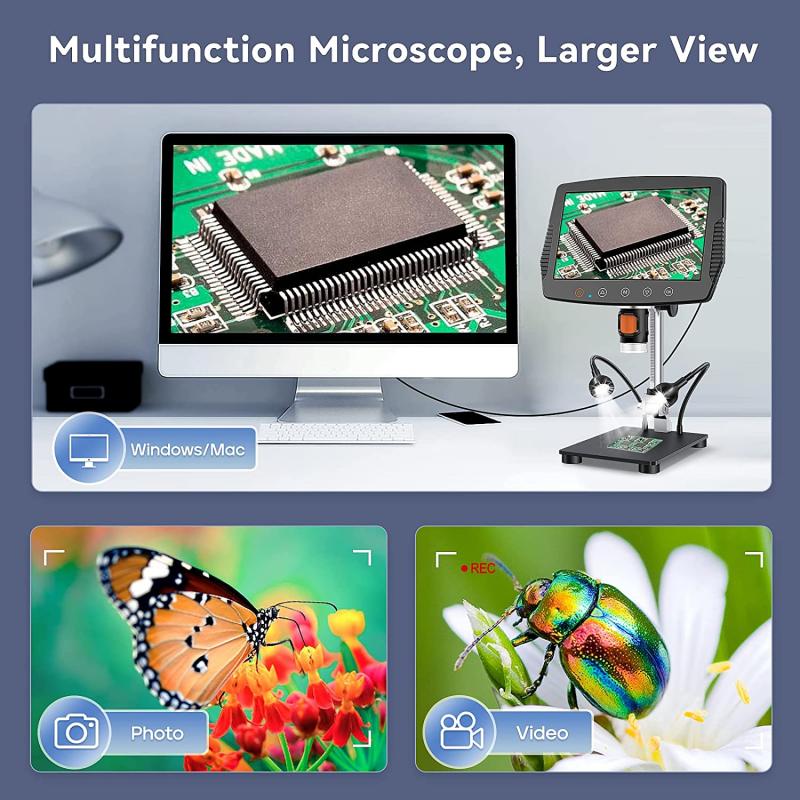
Yes, bacteria can be seen with a microscope. In fact, the invention of the microscope in the 17th century allowed scientists to observe and study microorganisms for the first time. With the use of light microscopes, bacteria can be visualized at magnifications of up to 1000x. This allows scientists to observe the shape, size, and arrangement of bacterial cells, as well as their internal structures such as the cell wall, cytoplasm, and nucleoid.
In recent years, advances in microscopy technology have allowed for even greater visualization of bacterial structures. Electron microscopy, for example, uses beams of electrons to create highly detailed images of bacterial cells and their components. This has allowed scientists to study the ultrastructure of bacteria, including the structure of their cell walls, flagella, and pili.
Fluorescence microscopy is another technique that has revolutionized the study of bacteria. By labeling specific bacterial structures with fluorescent dyes, scientists can observe the dynamics of bacterial processes such as cell division and protein localization in real-time. This has led to a greater understanding of the complex interactions between bacteria and their environment.
Overall, the ability to visualize bacterial structures has been crucial in advancing our understanding of these microorganisms and their role in the world around us.













There are no comments for this blog.