Can You See Molecules With A Microscope ?
Yes, it is possible to see molecules with a microscope, but it depends on the type of microscope being used. Traditional light microscopes are not powerful enough to see individual molecules, as they have a resolution limit of around 200 nanometers. However, advanced techniques such as scanning tunneling microscopy (STM) and atomic force microscopy (AFM) can visualize individual atoms and molecules. These techniques work by scanning a tiny probe over the surface of a sample, measuring the forces between the probe and the molecules. The resulting data is then used to create a high-resolution image of the sample, revealing the positions of individual molecules.
1、 Types of Microscopes

There are several types of microscopes available, each with its own unique features and capabilities. These include optical microscopes, electron microscopes, and scanning probe microscopes.
Optical microscopes use visible light to magnify objects, and are commonly used in biology and medicine to study cells and tissues. However, they have limitations in terms of resolution and cannot see molecules directly.
Electron microscopes, on the other hand, use beams of electrons to create highly detailed images of objects at the atomic and molecular level. This allows scientists to see individual molecules and study their structure and function. However, electron microscopes are expensive and require specialized training to operate.
Scanning probe microscopes use a tiny probe to scan the surface of a sample and create a three-dimensional image. They can also be used to manipulate individual molecules and atoms, making them useful for nanotechnology research.
In recent years, advances in technology have led to the development of new types of microscopes, such as super-resolution microscopes that can see structures smaller than the diffraction limit of light. These new tools are helping scientists to better understand the complex world of molecules and atoms, and are opening up new avenues for research in fields such as medicine, materials science, and nanotechnology.
2、 - Optical

Optical microscopes are commonly used in laboratories to observe biological specimens, cells, and tissues. However, the question of whether we can see molecules with an optical microscope is a bit more complicated.
The short answer is no, we cannot see individual molecules with an optical microscope. This is because the resolution of an optical microscope is limited by the wavelength of light used to illuminate the sample. The smallest object that can be resolved by an optical microscope is about 200 nanometers, which is much larger than most molecules.
However, recent advances in microscopy techniques have allowed scientists to visualize individual molecules using fluorescence microscopy. This technique involves labeling molecules with fluorescent dyes or proteins that emit light when excited by a specific wavelength of light. By using specialized microscopes and imaging software, scientists can track the movement and interactions of individual molecules in real-time.
Another technique that has been developed to visualize molecules is cryo-electron microscopy (cryo-EM). This technique involves freezing samples at extremely low temperatures and imaging them with an electron microscope. Cryo-EM has revolutionized the field of structural biology, allowing scientists to determine the 3D structures of large biomolecules such as proteins and viruses at near-atomic resolution.
In summary, while optical microscopes cannot directly visualize individual molecules, advances in microscopy techniques such as fluorescence microscopy and cryo-EM have allowed scientists to study the behavior and structure of molecules in unprecedented detail.
3、 - Electron
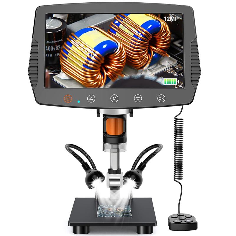
Electron microscopes are powerful tools that use beams of electrons to create high-resolution images of objects. Unlike traditional light microscopes, electron microscopes can magnify objects up to 10 million times, allowing scientists to see structures that are too small to be seen with traditional microscopes.
With an electron microscope, it is possible to see individual atoms and molecules. However, it is important to note that the process of imaging molecules with an electron microscope can be challenging. Molecules are not visible in their natural state, and they must be prepared in a way that allows them to be imaged.
One technique that is commonly used to image molecules with an electron microscope is called cryo-electron microscopy (cryo-EM). This technique involves freezing molecules in a thin layer of ice and then imaging them with an electron microscope. Cryo-EM has revolutionized the field of structural biology, allowing scientists to determine the structures of complex molecules such as proteins and viruses.
In summary, while it is possible to see molecules with an electron microscope, it requires specialized techniques such as cryo-EM. These techniques have opened up new avenues for research in fields such as structural biology and materials science.
4、 - Scanning Probe

Scanning probe microscopy (SPM) is a family of techniques that allows the imaging and manipulation of matter at the nanoscale. The most common types of SPM are atomic force microscopy (AFM) and scanning tunneling microscopy (STM). These techniques use a sharp probe that is scanned over the surface of a sample to measure its properties with high resolution.
While SPM can provide detailed information about the structure and properties of materials at the atomic and molecular scale, it is important to note that it does not directly visualize individual molecules. This is because the probes used in SPM are typically much larger than individual molecules, and they interact with the sample in a complex way that depends on the properties of the probe and the sample.
However, recent advances in SPM have allowed researchers to indirectly visualize the positions of individual molecules by using functionalized probes that can selectively bind to specific molecules on a surface. This approach, known as scanning probe lithography, has been used to create complex patterns and structures at the nanoscale.
Overall, while SPM cannot directly visualize individual molecules, it remains a powerful tool for studying the properties of materials at the nanoscale and for manipulating matter with high precision.











There are no comments for this blog.