Endoscopic Ultrasound Eus What Is It ?
Endoscopic ultrasound (EUS) is a medical procedure that combines endoscopy and ultrasound imaging. It involves the use of a specialized endoscope equipped with an ultrasound probe at its tip. The endoscope is inserted into the body through the mouth or rectum to visualize and obtain detailed images of the gastrointestinal tract and nearby organs. EUS allows for the evaluation of the walls of the digestive tract, as well as the surrounding lymph nodes, blood vessels, and other structures. It is commonly used for diagnosing and staging various gastrointestinal conditions, such as tumors, cysts, and inflammatory diseases. EUS can also be used to guide fine needle aspiration (FNA) or biopsy procedures, where a small tissue sample is obtained for further analysis. This minimally invasive technique provides valuable information for the diagnosis and management of gastrointestinal disorders.
1、 Definition and Purpose of Endoscopic Ultrasound (EUS)
Endoscopic ultrasound (EUS) is a minimally invasive procedure that combines endoscopy and ultrasound imaging to provide detailed images of the digestive tract and surrounding organs. It involves the use of a flexible endoscope, which is a long, thin tube with a light and camera at the end, and an ultrasound probe that is attached to the endoscope.
During an EUS procedure, the endoscope is inserted through the mouth or anus and advanced into the esophagus, stomach, or rectum, depending on the area being examined. The ultrasound probe emits high-frequency sound waves that create detailed images of the digestive tract and nearby structures, such as the pancreas, liver, and lymph nodes. These images can help diagnose and stage various gastrointestinal conditions, including cancers, cysts, and inflammatory diseases.
EUS has several advantages over other imaging techniques. It allows for a closer and more detailed examination of the digestive tract and surrounding organs, as the ultrasound probe is positioned directly next to the area of interest. This enables the detection of small lesions that may not be visible with other imaging modalities. EUS also allows for the collection of tissue samples (biopsies) for further analysis, which can aid in the diagnosis and treatment planning.
In recent years, EUS has seen advancements in technology, such as the development of high-frequency ultrasound probes and the integration of advanced imaging techniques like elastography and contrast-enhanced imaging. These advancements have further improved the accuracy and diagnostic capabilities of EUS.
Overall, EUS is a valuable tool in the diagnosis and management of gastrointestinal conditions. It provides detailed imaging and the ability to obtain tissue samples, making it an essential procedure in the field of gastroenterology and oncology.
2、 EUS Procedure and Equipment
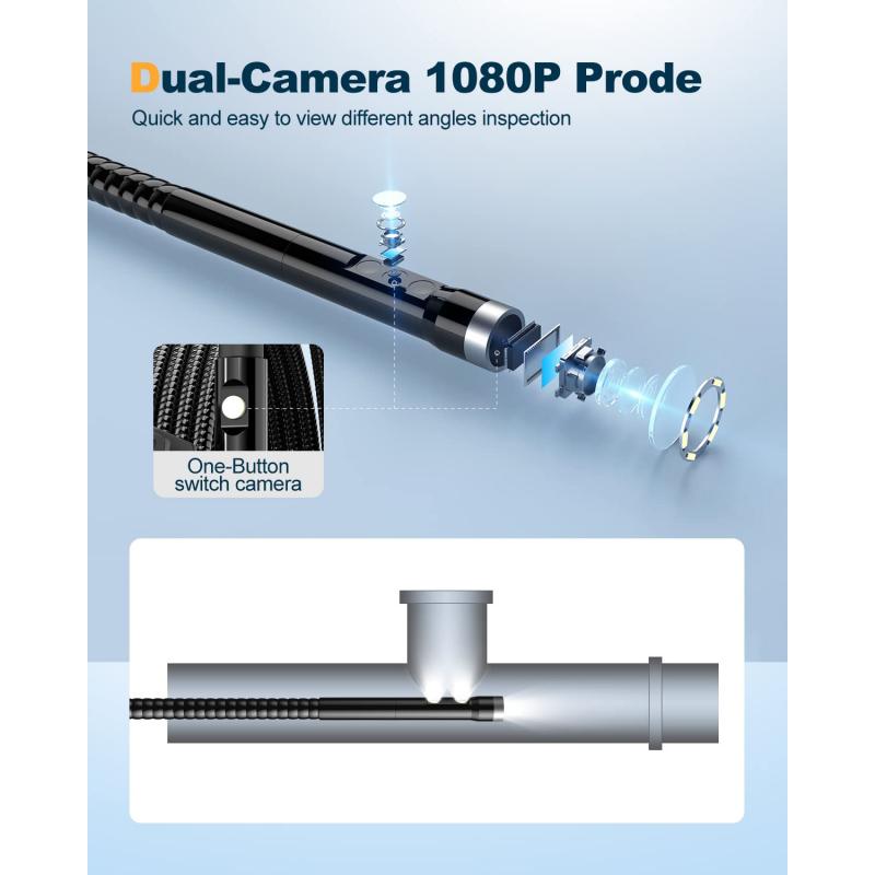
Endoscopic ultrasound (EUS) is a minimally invasive procedure that combines endoscopy and ultrasound imaging to visualize and evaluate the digestive tract and surrounding organs. It involves the use of a flexible endoscope equipped with an ultrasound probe at its tip, which allows for detailed imaging of the gastrointestinal (GI) tract and adjacent structures.
During an EUS procedure, the endoscope is inserted through the mouth or anus and advanced into the esophagus, stomach, or rectum, depending on the area being examined. The ultrasound probe emits high-frequency sound waves that create detailed images of the GI tract layers and nearby organs, such as the liver, pancreas, and lymph nodes. These images provide valuable information about the size, location, and characteristics of tumors, cysts, and other abnormalities.
EUS has become an essential tool in the diagnosis and staging of GI cancers, as it allows for accurate assessment of tumor depth, involvement of nearby structures, and presence of metastasis. It is also used to guide fine needle aspiration (FNA) biopsies, where a small needle is inserted through the endoscope to obtain tissue samples for further analysis.
In recent years, advancements in EUS technology have further improved its diagnostic capabilities. High-frequency ultrasound probes provide higher resolution images, allowing for better visualization of small lesions and early-stage tumors. Additionally, the development of therapeutic EUS techniques, such as EUS-guided ablation and EUS-guided drainage of fluid collections, has expanded the range of conditions that can be treated using this procedure.
Overall, EUS is a valuable tool in the field of gastroenterology and oncology, providing clinicians with detailed imaging and the ability to perform targeted biopsies and therapeutic interventions. Its non-invasive nature and high diagnostic accuracy make it a preferred choice for evaluating a wide range of GI conditions.

3、 Applications of EUS in Gastroenterology
Endoscopic ultrasound (EUS) is a minimally invasive procedure that combines endoscopy and ultrasound to obtain detailed images of the gastrointestinal tract and surrounding structures. It involves the use of a specialized endoscope equipped with an ultrasound probe at its tip, which allows for high-resolution imaging of the digestive system.
EUS has a wide range of applications in the field of gastroenterology. One of its primary uses is in the evaluation and staging of gastrointestinal cancers. EUS can accurately assess the depth of tumor invasion, involvement of nearby lymph nodes, and presence of distant metastases, aiding in the selection of appropriate treatment strategies. It is particularly useful in the staging of esophageal, gastric, pancreatic, and rectal cancers.
In addition to cancer staging, EUS is also valuable in the diagnosis and management of various gastrointestinal conditions. It can help identify the cause of unexplained abdominal pain, evaluate pancreatic and biliary disorders, detect gallstones, assess the severity of chronic pancreatitis, and guide the placement of drainage tubes or needles for therapeutic interventions.
Furthermore, EUS has emerged as a valuable tool for obtaining tissue samples for pathological analysis. Fine-needle aspiration (FNA) or fine-needle biopsy (FNB) can be performed during EUS to obtain samples from suspicious lesions or lymph nodes. This allows for accurate diagnosis of malignancies and aids in the selection of appropriate treatment options.
The latest point of view regarding EUS is the development of advanced imaging techniques and technologies. These include the use of contrast-enhanced EUS, which improves the visualization of blood vessels and enhances the detection of small lesions. Additionally, elastography, a technique that assesses tissue stiffness, has shown promise in differentiating benign from malignant lesions.
In conclusion, EUS is a versatile and valuable tool in gastroenterology. Its applications range from cancer staging to the diagnosis and management of various gastrointestinal conditions. With the advent of advanced imaging techniques, EUS continues to evolve and improve, providing clinicians with more accurate and detailed information for optimal patient care.

4、 EUS in the Diagnosis of Pancreatic Diseases
Endoscopic ultrasound (EUS) is a minimally invasive procedure that combines endoscopy and ultrasound imaging to visualize and evaluate the gastrointestinal tract and surrounding structures. It involves the use of a flexible endoscope with an ultrasound probe attached to its tip, which allows for high-resolution imaging of the pancreas and other organs in the abdomen.
EUS has emerged as a valuable tool in the diagnosis of pancreatic diseases. It provides detailed images of the pancreas, allowing for the detection and characterization of various conditions such as pancreatic tumors, cysts, and chronic pancreatitis. EUS can also be used to guide fine needle aspiration (FNA) biopsies, where a small tissue sample is obtained for further analysis.
One of the major advantages of EUS is its ability to provide more accurate and detailed information compared to other imaging modalities. It can detect small lesions that may not be visible on other imaging tests like CT scans or MRI. EUS also allows for real-time imaging, which enables the physician to visualize the pancreas and surrounding structures from different angles and obtain more precise measurements.
In recent years, there have been advancements in EUS technology, such as the development of high-frequency ultrasound probes and the addition of contrast-enhanced imaging techniques. These advancements have further improved the diagnostic capabilities of EUS, particularly in the evaluation of pancreatic tumors. Contrast-enhanced EUS can help differentiate between benign and malignant lesions based on their vascularity patterns.
Overall, EUS has become an essential tool in the diagnosis and management of pancreatic diseases. It offers a safe and effective means of obtaining detailed imaging and tissue samples for accurate diagnosis. As technology continues to advance, EUS is likely to play an even more significant role in the early detection and treatment of pancreatic diseases.
