Fluorescent Microscope How It Works ?
A fluorescent microscope is a type of microscope that uses fluorescence to generate an image. It works by exciting fluorescent molecules in a sample with a specific wavelength of light, causing them to emit light at a longer wavelength. This emitted light is then detected by the microscope and used to create an image.
To achieve this, the microscope uses a light source, such as a mercury or xenon lamp, to produce a beam of light that is filtered to a specific wavelength. This light is then directed onto the sample, which has been labeled with fluorescent molecules, such as dyes or proteins.
When the fluorescent molecules are excited by the light, they emit light at a longer wavelength, which is then captured by the microscope's detector. The detector then converts this light into an electrical signal, which is used to create an image of the sample.
Fluorescent microscopy is widely used in biological research, as it allows scientists to visualize specific molecules or structures within cells and tissues. It is also used in medical diagnostics, such as detecting cancer cells or infectious agents.
1、 Excitation light source

Fluorescent microscope how it works: Excitation light source
The excitation light source is a critical component of a fluorescent microscope. It provides the energy required to excite the fluorescent molecules in the sample, causing them to emit light of a different wavelength. The excitation light source can be a lamp, laser, or LED, depending on the specific application.
In a traditional fluorescent microscope, a mercury arc lamp is used as the excitation light source. The lamp emits a broad spectrum of light, which is filtered to select the appropriate wavelength for excitation of the fluorescent molecules. The filtered light is then directed onto the sample through the objective lens.
In recent years, LED light sources have become increasingly popular for use in fluorescent microscopy. LEDs offer several advantages over traditional lamps, including longer lifetimes, lower power consumption, and greater stability. Additionally, LEDs can be tuned to emit specific wavelengths of light, allowing for more precise excitation of fluorescent molecules.
Another emerging technology in the field of fluorescent microscopy is super-resolution microscopy. This technique allows for imaging at resolutions beyond the diffraction limit of light, enabling researchers to visualize structures and processes at the molecular level. One approach to super-resolution microscopy is stimulated emission depletion (STED) microscopy, which uses a laser to excite fluorescent molecules in a small region of the sample. A second laser is then used to de-excite the molecules, resulting in a highly localized fluorescent signal. STED microscopy has been used to image structures as small as 20 nanometers, providing unprecedented insights into the organization and function of biological systems.
In summary, the excitation light source is a critical component of a fluorescent microscope, providing the energy required to excite fluorescent molecules in the sample. Traditional mercury arc lamps are commonly used, but LED light sources are becoming increasingly popular due to their advantages in terms of lifetime, power consumption, and stability. Emerging technologies such as super-resolution microscopy are also pushing the boundaries of what is possible with fluorescent microscopy.
2、 Excitation filter
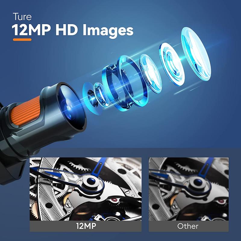
Fluorescent microscope how it works:
A fluorescent microscope is an optical microscope that uses fluorescence to generate an image. It works by exciting fluorescent molecules with a specific wavelength of light and then detecting the emitted light. The excitation filter is a critical component of the fluorescent microscope. It is a filter that allows only the excitation wavelength to pass through to the sample, while blocking all other wavelengths of light.
When the excitation filter is placed in front of the light source, it allows only the specific wavelength of light to pass through to the sample. This light then excites the fluorescent molecules in the sample, causing them to emit light at a longer wavelength. The emitted light is then detected by the microscope's detector, which produces an image of the sample.
The excitation filter is designed to match the excitation wavelength of the fluorescent molecule being used. This ensures that only the desired molecules are excited and detected, while other molecules in the sample are not excited and do not contribute to the image.
Recent advancements in fluorescent microscopy have led to the development of super-resolution microscopy techniques, such as stimulated emission depletion (STED) microscopy and structured illumination microscopy (SIM). These techniques use specialized excitation filters and other components to achieve resolutions beyond the diffraction limit of light, allowing for the visualization of structures and processes at the nanoscale level.
3、 Dichroic mirror
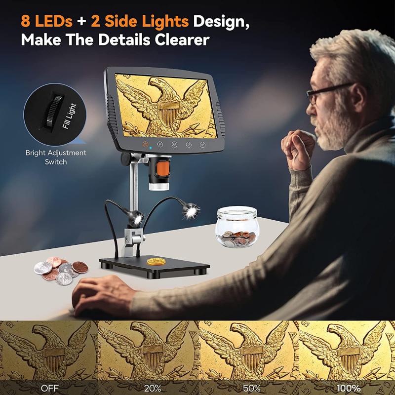
Dichroic mirror is a key component of a fluorescent microscope that allows for the separation of excitation and emission light. It is a specialized mirror that reflects light of a certain wavelength while allowing light of other wavelengths to pass through. In a fluorescent microscope, the dichroic mirror is placed at a 45-degree angle between the objective lens and the light source.
When light from the excitation source (usually a high-intensity lamp or laser) passes through the dichroic mirror, it is reflected towards the sample. The sample absorbs the excitation light and emits light of a longer wavelength, which is then collected by the objective lens and passes through the dichroic mirror. However, the dichroic mirror reflects the emitted light towards the detector, while allowing any remaining excitation light to pass through and be absorbed by a filter.
The use of a dichroic mirror in a fluorescent microscope allows for the separation of excitation and emission light, which is crucial for obtaining clear and accurate images. It also allows for the use of multiple fluorophores in a single sample, as each fluorophore can be excited by a different wavelength of light and detected separately.
Recent advancements in technology have led to the development of more advanced dichroic mirrors, such as those with higher reflectivity and better spectral separation. These improvements have allowed for more precise and sensitive imaging, particularly in live-cell imaging and super-resolution microscopy.
4、 Emission filter
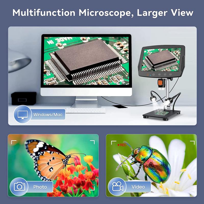
Fluorescent microscope how it works:
A fluorescent microscope is an optical microscope that uses fluorescence to generate an image. It works by exciting fluorescent molecules with a specific wavelength of light, causing them to emit light of a different wavelength. This emitted light is then detected by the microscope and used to create an image.
One important component of a fluorescent microscope is the emission filter. This filter is placed between the sample and the detector and is designed to block any excitation light from reaching the detector while allowing the emitted light to pass through. This is important because without the emission filter, the excitation light would overwhelm the emitted light, making it difficult to see the fluorescent signal.
The emission filter is typically made of a thin layer of material that has been coated with a series of interference filters. These filters are designed to reflect light of certain wavelengths while allowing light of other wavelengths to pass through. By carefully selecting the thickness and composition of these filters, it is possible to create a filter that blocks the excitation light while allowing the emitted light to pass through.
In recent years, there have been several advances in the design of emission filters. For example, some filters now use multiple layers of interference filters to create a more precise wavelength cutoff. Additionally, some filters are now designed to be tunable, allowing researchers to adjust the wavelength cutoff to better match the fluorescent molecules being used.
Overall, the emission filter is a critical component of a fluorescent microscope, allowing researchers to see the fluorescent signal without interference from the excitation light. As technology continues to advance, it is likely that we will see further improvements in the design and performance of these filters.


