How A Compound Light Microscope Works ?
A compound light microscope works by using two lenses to magnify an object. The objective lens is located near the specimen and produces a magnified image that is further magnified by the eyepiece lens. Light passes through the specimen and is refracted by the objective lens, which produces a real, inverted image. The eyepiece lens then magnifies this image and presents it to the viewer's eye. The total magnification is the product of the magnification of the objective lens and the eyepiece lens. The microscope also contains a light source that illuminates the specimen, and various adjustments can be made to focus the image and adjust the amount of light.
1、 Basic components of a compound light microscope
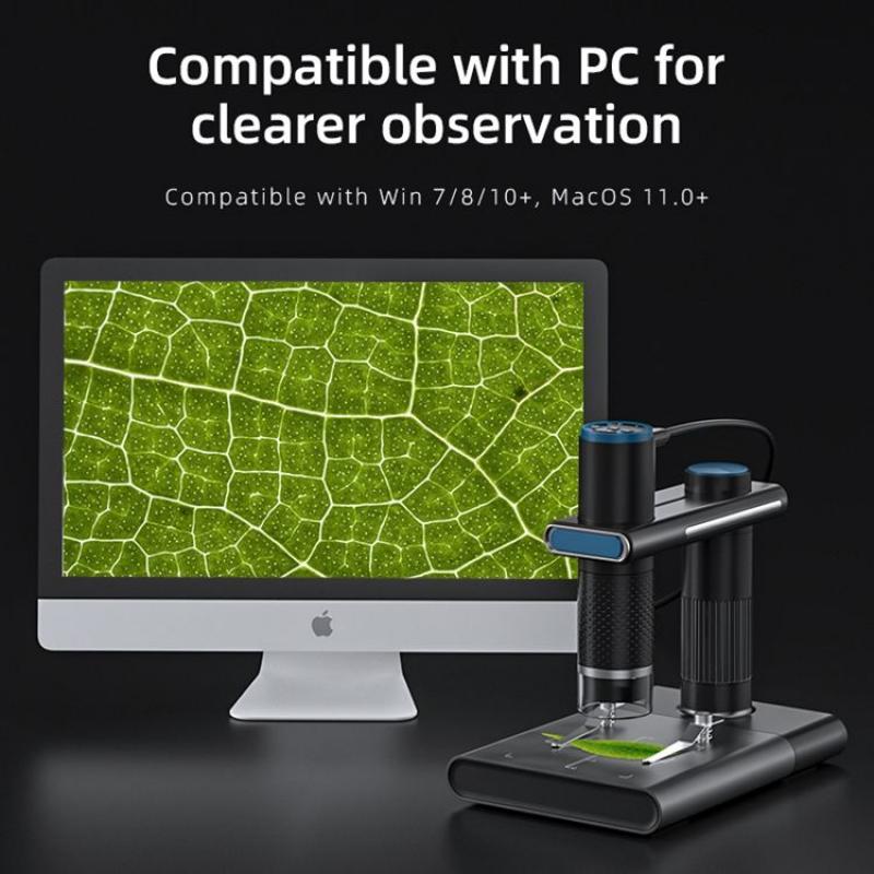
Basic components of a compound light microscope include an eyepiece, objective lenses, a stage, a light source, and a focusing mechanism. The eyepiece is the lens that the viewer looks through to observe the specimen. The objective lenses are located on a rotating turret and provide different levels of magnification. The stage is where the specimen is placed and can be moved up and down to adjust the focus. The light source illuminates the specimen, allowing it to be seen more clearly. The focusing mechanism moves the stage up and down to bring the specimen into focus.
A compound light microscope works by using a series of lenses to magnify the specimen. The light source shines light through the specimen, which is then magnified by the objective lenses. The magnified image is then further magnified by the eyepiece, which allows the viewer to see the specimen in greater detail. The focusing mechanism is used to adjust the distance between the objective lenses and the specimen, which changes the level of magnification and allows the viewer to bring the specimen into focus.
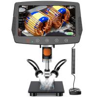
In recent years, advancements in technology have led to the development of digital microscopes, which use a camera to capture images of the specimen and display them on a computer screen. These microscopes allow for greater accuracy and precision in observation and analysis, as well as the ability to easily share and store images. Additionally, some microscopes now use fluorescent dyes to highlight specific structures within the specimen, allowing for even greater detail and specificity in observation.
2、 Principles of light refraction and magnification

A compound light microscope works by using a series of lenses to magnify an object. The principles of light refraction and magnification are key to its operation. When light passes through a lens, it is refracted or bent, causing the image to appear larger. The magnification of the image is determined by the power of the lens, which is a measure of its ability to bend light.
In a compound light microscope, there are two lenses: the objective lens and the eyepiece lens. The objective lens is located near the specimen and is responsible for magnifying the image. The eyepiece lens is located near the eye and further magnifies the image produced by the objective lens.
The light source in a compound light microscope illuminates the specimen, allowing it to be seen. The light passes through the objective lens and is refracted, producing a magnified image. The image is then further magnified by the eyepiece lens, allowing it to be seen by the observer.
Recent advancements in technology have allowed for the development of digital compound microscopes, which use a camera to capture images of the specimen. These images can then be viewed on a computer screen, allowing for easier sharing and analysis of data.
In conclusion, the principles of light refraction and magnification are essential to the operation of a compound light microscope. By using a series of lenses to magnify the image, it allows for the observation of small objects and structures that would otherwise be invisible to the naked eye. The latest advancements in technology have further improved the capabilities of the compound microscope, making it an invaluable tool in scientific research and discovery.
3、 Types of lenses used in a compound light microscope

How a compound light microscope works:
A compound light microscope works by using two lenses to magnify an object. The first lens, called the objective lens, is located near the specimen and magnifies the image. The second lens, called the eyepiece or ocular lens, is located near the eye and further magnifies the image. The combination of these two lenses allows for a much greater magnification than either lens could achieve on its own.
Light from a source, such as a bulb, passes through the specimen and is then focused by the objective lens. The image is then magnified and inverted. The light then passes through the eyepiece lens, which further magnifies the image and corrects the inversion. The final image is then viewed by the observer.
Types of lenses used in a compound light microscope:
There are several types of lenses used in a compound light microscope, including achromatic, apochromatic, and plan-achromatic lenses. Achromatic lenses are the most common and are made from two types of glass with different refractive indices. This helps to reduce chromatic aberration, which is when different colors of light are focused at different points, causing a blurry image.
Apochromatic lenses are made from three types of glass and are even better at reducing chromatic aberration. They are more expensive than achromatic lenses but provide a higher quality image.
Plan-achromatic lenses are similar to apochromatic lenses but also correct for spherical aberration, which is when light is focused at different points on a curved surface, causing distortion. These lenses are the most expensive but provide the highest quality image.
In recent years, there has been a trend towards using digital microscopes, which use a camera to capture images instead of an eyepiece. These microscopes can provide higher resolution images and allow for easier sharing and analysis of data.
4、 Adjusting focus and resolving power

A compound light microscope works by using a series of lenses to magnify an object. The lenses are arranged in a specific way to produce a magnified image that can be viewed through the eyepiece. The microscope has two lenses, the objective lens and the eyepiece lens. The objective lens is located near the specimen and produces a magnified image that is then further magnified by the eyepiece lens.
Adjusting focus is an important aspect of using a compound light microscope. The focus is adjusted by moving the objective lens closer or further away from the specimen. This allows the user to bring the specimen into sharp focus and see the details of the object being viewed. The resolving power of the microscope is also important. This refers to the ability of the microscope to distinguish between two closely spaced objects. The resolving power is determined by the wavelength of light used and the numerical aperture of the lens.
Recent advancements in technology have led to the development of digital microscopes. These microscopes use digital cameras to capture images of the specimen and display them on a computer screen. This allows for easier sharing of images and the ability to manipulate the images for further analysis. Additionally, some digital microscopes use fluorescence to highlight specific structures within the specimen, allowing for greater detail and understanding of the object being viewed.
In conclusion, a compound light microscope works by using lenses to magnify an object, with adjusting focus and resolving power being important aspects of its use. Advancements in technology have led to the development of digital microscopes, which offer new capabilities for viewing and analyzing specimens.









There are no comments for this blog.