How An Endoscope Works ?
An endoscope is a medical device used to examine the inside of the body. It consists of a long, thin, flexible tube with a light and camera at the end. The endoscope is inserted into the body through a natural opening or a small incision. The camera sends images to a monitor, allowing the doctor to see inside the body.
The endoscope is controlled by the doctor, who can move it around to get a better view of the area being examined. Some endoscopes also have tools attached to them, such as forceps or scissors, which can be used to take tissue samples or perform minor surgeries.
The endoscope is typically used to examine the digestive tract, including the esophagus, stomach, and intestines. It can also be used to examine other parts of the body, such as the lungs, bladder, and joints. The procedure is usually done under sedation or anesthesia to minimize discomfort for the patient.
1、 Optical System
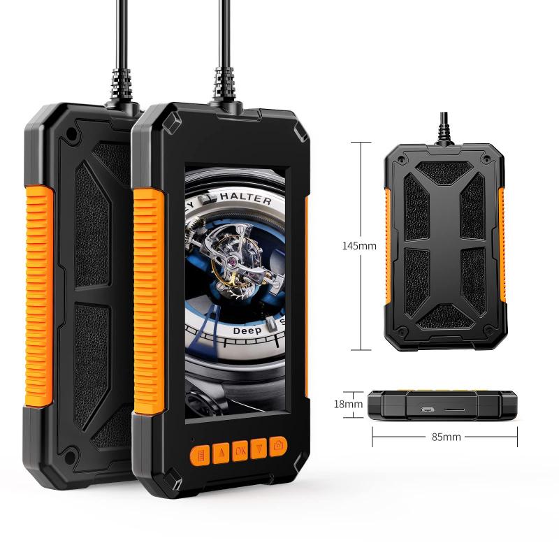
An endoscope is a medical device that is used to examine the internal organs and tissues of the human body. It consists of a long, thin, flexible tube with a camera and light source at the end. The camera captures images of the internal organs and tissues, which are then transmitted to a monitor for viewing by the medical professional.
The optical system is a critical component of an endoscope. It consists of a lens system that focuses the light from the light source onto the area being examined. The lens system also captures the reflected light from the area being examined and transmits it to the camera. The camera then converts the light into an electronic signal that is transmitted to the monitor for viewing.
The latest point of view on the optical system of an endoscope is the use of high-definition cameras and advanced lens systems. These advancements have greatly improved the quality of the images captured by the endoscope, making it easier for medical professionals to diagnose and treat various medical conditions.
In addition, some endoscopes now come equipped with advanced imaging technologies such as narrow-band imaging and fluorescence imaging. These technologies enhance the contrast and detail of the images captured by the endoscope, making it easier to detect abnormalities and diagnose medical conditions.
Overall, the optical system is a critical component of an endoscope, and advancements in this area have greatly improved the quality of medical care provided to patients.
2、 Light Source
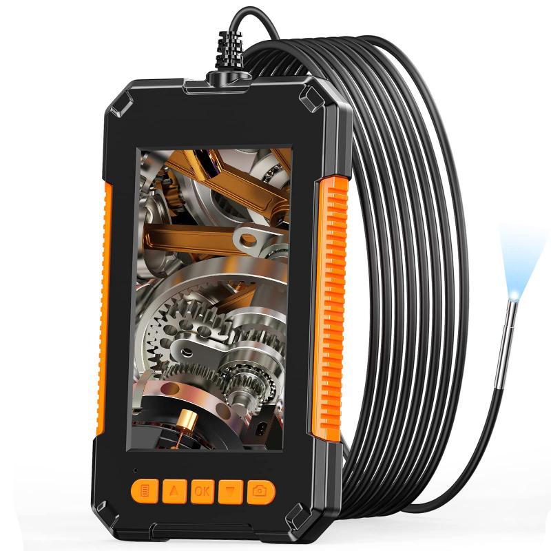
An endoscope is a medical device that is used to examine the inside of the body. It consists of a long, thin tube with a camera and light source at the end. The light source is an essential component of the endoscope as it illuminates the area being examined, allowing the camera to capture clear images.
The light source in an endoscope is typically a high-intensity LED or halogen bulb. The light is transmitted through a fiber optic cable that runs through the length of the endoscope. The fiber optic cable is made up of thousands of tiny glass fibers that are bundled together. When the light source is turned on, the light travels through the fiber optic cable and is emitted from the end of the endoscope.
The latest point of view on endoscope technology is the use of wireless endoscopes. These endoscopes do not require a fiber optic cable to transmit the light. Instead, they use a small camera and LED light source that are attached to the end of a flexible tube. The images captured by the camera are transmitted wirelessly to a monitor, allowing the doctor to view the inside of the body in real-time.
In conclusion, the light source is a crucial component of an endoscope as it provides the illumination necessary for clear imaging. The latest advancements in endoscope technology have led to the development of wireless endoscopes, which offer greater flexibility and ease of use.
3、 Fiber Optic Cable
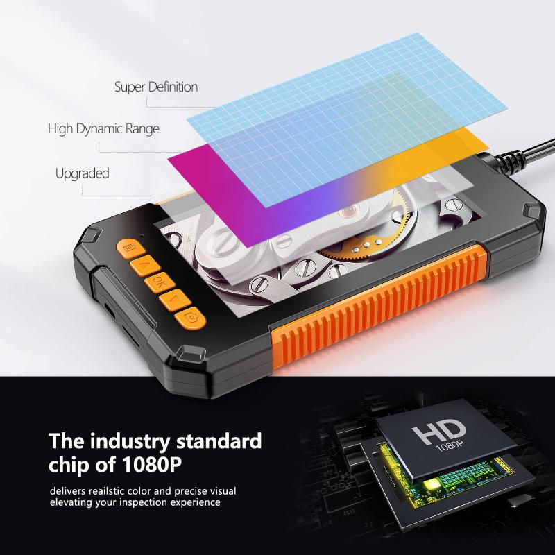
An endoscope is a medical device that is used to examine the inside of the body. It consists of a long, thin tube with a camera and light source at the end. The camera captures images of the inside of the body and sends them to a monitor for the doctor to view. One of the key components of an endoscope is the fiber optic cable.
A fiber optic cable is a thin, flexible cable made up of thousands of tiny glass or plastic fibers. These fibers are arranged in a bundle and are used to transmit light from one end of the cable to the other. In an endoscope, the fiber optic cable is used to transmit light from the light source at the end of the tube to the camera.
When light is shone into one end of the fiber optic cable, it travels down the length of the cable by bouncing off the walls of the individual fibers. This process is known as total internal reflection. The light emerges from the other end of the cable and illuminates the area being examined by the camera.
The use of fiber optic cables in endoscopes has revolutionized the field of medical imaging. They allow doctors to see inside the body without the need for invasive surgery. The latest advancements in fiber optic technology have led to the development of smaller, more flexible endoscopes that can reach even the most hard-to-reach areas of the body.
In conclusion, the fiber optic cable is a crucial component of an endoscope. It allows doctors to see inside the body and diagnose and treat a wide range of medical conditions. The latest advancements in fiber optic technology have made endoscopes even more effective and versatile, and they continue to play a vital role in modern medicine.
4、 Camera System
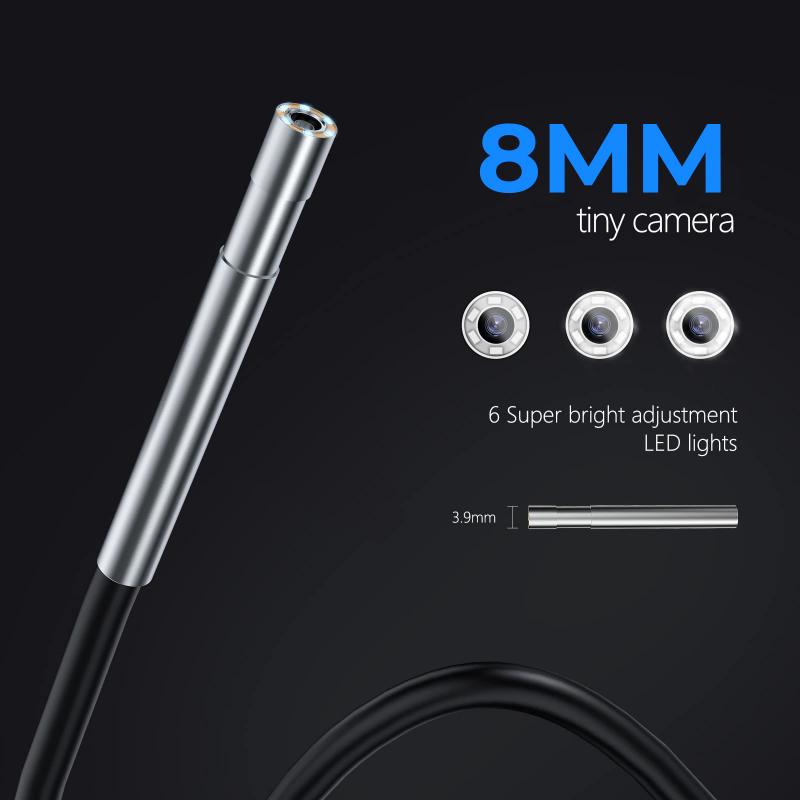
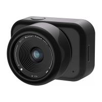
An endoscope is a medical device that is used to examine the inside of the body. It is a long, thin, flexible tube that has a camera and a light source at the end. The camera system is the most important part of the endoscope, as it allows doctors to see inside the body without making any incisions.
The camera system in an endoscope works by using a small camera that is attached to the end of the tube. The camera captures images of the inside of the body and sends them to a monitor, where the doctor can view them in real-time. The light source at the end of the tube illuminates the area being examined, making it easier for the camera to capture clear images.
The latest point of view in endoscopy is the use of high-definition cameras, which provide even clearer images than traditional cameras. These cameras have a higher resolution and can capture more detail, making it easier for doctors to identify any abnormalities or issues.
In addition to the camera system, endoscopes may also have other tools attached to them, such as forceps or scissors, which can be used to take tissue samples or perform minor procedures.
Overall, the camera system is a crucial component of an endoscope, allowing doctors to see inside the body and diagnose and treat a wide range of medical conditions. With the latest advancements in technology, endoscopes are becoming even more effective and precise, improving patient outcomes and reducing the need for invasive procedures.












There are no comments for this blog.