How Big Is An Electron Microscope ?
The size of an electron microscope can vary depending on the specific model and design. Generally, electron microscopes are larger than optical microscopes due to the complex technology involved. They can range in size from a few meters in length to several meters in height. The main components of an electron microscope, such as the electron source, electron lenses, and detectors, contribute to its overall size. Additionally, electron microscopes require a dedicated space with proper infrastructure, including a stable environment, electrical supply, and cooling systems.
1、 Magnification capabilities of an electron microscope
The size of an electron microscope can vary depending on the specific model and type. Generally, electron microscopes are larger than optical microscopes due to the complex technology involved. They consist of several components, including a vacuum chamber, electron gun, electromagnetic lenses, and detectors. The overall size can range from a few meters in length for transmission electron microscopes (TEMs) to smaller, more compact designs for scanning electron microscopes (SEMs).
Regarding the magnification capabilities of an electron microscope, they are significantly higher than those of optical microscopes. Electron microscopes use a beam of electrons instead of light to visualize samples, allowing for much greater resolution and magnification. TEMs can achieve magnifications up to several million times, enabling the observation of atomic structures and individual molecules. SEMs, on the other hand, typically have lower magnification capabilities, ranging from a few hundred to a few hundred thousand times. However, SEMs excel in providing detailed surface information and three-dimensional imaging.
It is important to note that advancements in technology continue to push the boundaries of electron microscope capabilities. For instance, recent developments in aberration correction techniques have significantly improved the resolution and image quality of electron microscopes. Additionally, the integration of advanced detectors and imaging software has enhanced the efficiency and versatility of these instruments.
In summary, electron microscopes are larger than optical microscopes and offer much higher magnification capabilities. They are essential tools in various scientific fields, including materials science, biology, and nanotechnology. Ongoing advancements in electron microscope technology continue to expand their capabilities, enabling researchers to explore the microscopic world with unprecedented detail and precision.

2、 Resolution of an electron microscope
The size of an electron microscope can vary depending on the specific type and model. Generally, electron microscopes are larger than optical microscopes due to the complex technology involved. They can range in size from a few meters in length to several meters in height. The size is primarily determined by the need to accommodate the electron beam column, vacuum chamber, and other components necessary for the microscope's operation.
On the other hand, the resolution of an electron microscope refers to its ability to distinguish between two closely spaced objects. Electron microscopes have significantly higher resolution compared to optical microscopes due to the shorter wavelength of electrons. The resolution of an electron microscope is typically measured in nanometers (nm), and modern electron microscopes can achieve resolutions as low as 0.05 nm or even better.
It is important to note that the resolution of an electron microscope is not solely determined by its size. Other factors, such as the quality of the electron source, the stability of the microscope, and the design of the electron optics, also play a crucial role in achieving high-resolution imaging.
In recent years, advancements in electron microscopy techniques have led to even higher resolutions. For example, the development of aberration-corrected electron microscopy has allowed for atomic-scale imaging, enabling scientists to observe individual atoms and their arrangements within materials. This has opened up new possibilities for studying nanoscale structures and materials at an unprecedented level of detail.
Overall, the size of an electron microscope can vary, but its resolution is determined by various factors and can reach extremely high levels, allowing for detailed imaging at the atomic scale.

3、 Electron beam generation in an electron microscope
An electron microscope is a powerful scientific instrument used to observe objects at a very high resolution. It utilizes a beam of electrons instead of light to magnify and visualize the sample. The size of an electron microscope can vary depending on the type and purpose.
There are two main types of electron microscopes: transmission electron microscopes (TEM) and scanning electron microscopes (SEM). TEMs are used to study the internal structure of thin samples, while SEMs provide detailed surface information.
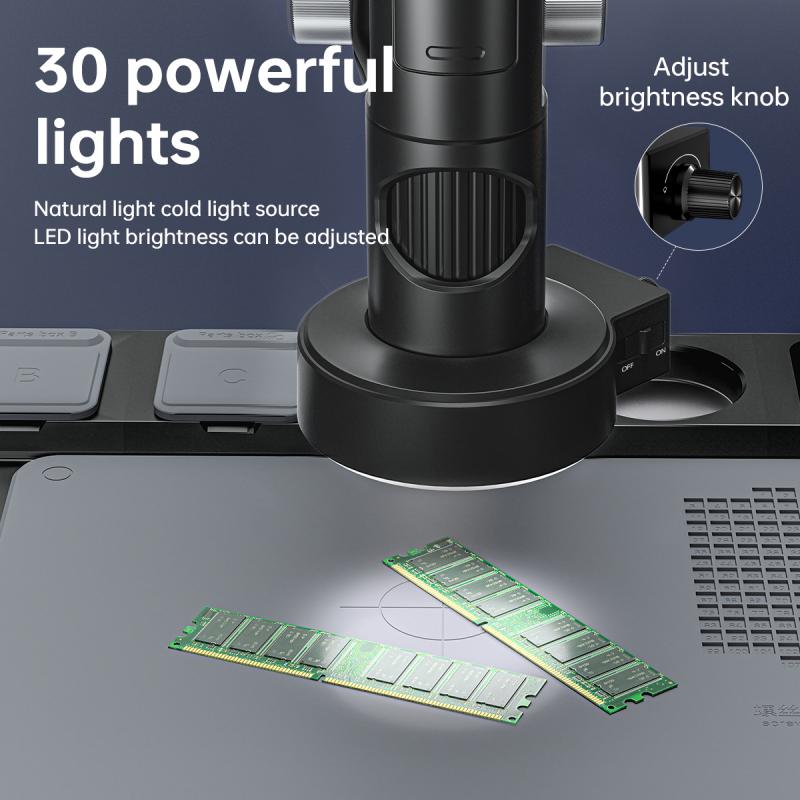
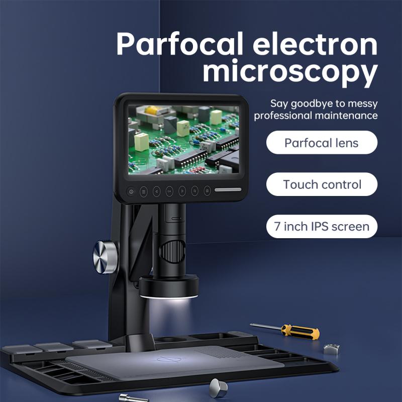
The size of an electron microscope can range from a small desktop model to a large, room-sized instrument. Desktop electron microscopes, also known as benchtop or tabletop microscopes, are compact and portable, making them suitable for smaller laboratories or fieldwork. These microscopes typically have a size of a few feet in height and width.
On the other hand, larger electron microscopes, especially those used in research institutions or industrial settings, can be quite massive. They can occupy an entire room and require specialized infrastructure to support their operation. These larger microscopes can have dimensions of several meters in height, width, and length.
In terms of electron beam generation, both TEMs and SEMs use similar principles. A high-voltage electron gun generates a beam of electrons, which is then focused and accelerated towards the sample. The electron beam interacts with the sample, and the resulting signals are detected and processed to create an image.
The latest advancements in electron microscopy have led to the development of more compact and efficient electron sources. For example, field-emission electron guns (FEG) have become increasingly popular due to their high brightness and stability. FEGs can produce a highly focused electron beam with a small spot size, enabling even higher resolution imaging.
In conclusion, the size of an electron microscope can vary depending on the type and purpose. From small desktop models to large room-sized instruments, electron microscopes are powerful tools for scientific research and industrial applications. The latest advancements in electron beam generation, such as field-emission electron guns, have further improved the resolution and performance of electron microscopes.

4、 Electron microscope imaging techniques and modes
An electron microscope is a powerful scientific instrument used to observe and analyze objects at the atomic and molecular level. It utilizes a beam of electrons instead of light to magnify and resolve the fine details of a specimen.
In terms of size, electron microscopes can vary depending on the specific model and purpose. The overall dimensions of an electron microscope can range from a few meters in length to a compact desktop size. The size is mainly determined by the complexity of the instrument and the required components for electron generation, focusing, and detection.
Electron microscope imaging techniques and modes have evolved significantly over the years, allowing for a wide range of applications. The most common imaging technique is called scanning electron microscopy (SEM), which provides high-resolution, three-dimensional images of the surface of a specimen. Transmission electron microscopy (TEM) is another widely used technique that allows for the visualization of internal structures of a specimen by passing electrons through it.
In recent years, advancements in electron microscopy have led to the development of new imaging modes and techniques. For example, cryo-electron microscopy (cryo-EM) has revolutionized the field of structural biology by enabling the visualization of biological macromolecules in their native state. This technique has been instrumental in determining the structures of complex proteins and viruses, leading to breakthroughs in drug discovery and understanding of disease mechanisms.
Furthermore, electron microscopy has also been combined with other imaging techniques, such as spectroscopy and tomography, to provide additional information about the chemical composition and three-dimensional structure of a specimen.
Overall, electron microscopes have become indispensable tools in various scientific disciplines, including materials science, nanotechnology, biology, and medicine. Their size and capabilities continue to evolve as technology advances, allowing researchers to explore the microscopic world with unprecedented detail and precision.


There are no comments for this blog.