How Do Conventional Light Microscopes Work ?
Conventional light microscopes work by using visible light to magnify and resolve small objects or structures. The light passes through the specimen and is refracted by the lenses of the microscope, which magnify the image and project it onto the eyepiece or camera. The lenses of the microscope are designed to bend the light in a way that allows the viewer to see a magnified image of the specimen. The amount of magnification depends on the objective lens used, which can be changed to increase or decrease the magnification. The resolution of the microscope is limited by the wavelength of visible light, which is around 400-700 nanometers. This means that structures smaller than this cannot be resolved by a conventional light microscope. To improve resolution, techniques such as staining or using specialized lenses can be used.
1、 Optical components and magnification
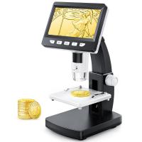
Conventional light microscopes work by using a series of optical components to magnify and focus light onto a specimen. The basic components of a light microscope include an objective lens, an eyepiece, and a light source. The objective lens is responsible for magnifying the specimen, while the eyepiece allows the viewer to see the magnified image. The light source provides illumination for the specimen.
When light passes through the objective lens, it is refracted and focused onto the specimen. The light then passes through the specimen and is refracted again as it passes through the objective lens. This creates a magnified image of the specimen that can be viewed through the eyepiece.
The magnification of a light microscope is determined by the combination of the objective lens and the eyepiece. The objective lens typically has a magnification of 4x, 10x, 40x, or 100x, while the eyepiece usually has a magnification of 10x. This means that the total magnification of a light microscope can range from 40x to 1000x.
While conventional light microscopes have been used for centuries, recent advancements in technology have allowed for the development of more advanced microscopy techniques. For example, confocal microscopy uses lasers to create a 3D image of a specimen, while super-resolution microscopy allows for the visualization of structures that are smaller than the diffraction limit of light. These techniques have revolutionized the field of microscopy and have allowed for new discoveries in biology and medicine.

2、 Illumination and contrast
Conventional light microscopes work by using a combination of illumination and contrast techniques to magnify and visualize small objects or structures. The microscope uses a light source, typically a bulb or LED, to illuminate the sample being observed. The light passes through a series of lenses, including the objective lens, which magnifies the image of the sample. The magnified image is then viewed through the eyepiece of the microscope.
In addition to illumination, contrast techniques are used to enhance the visibility of the sample. One common technique is staining, where a dye or chemical is applied to the sample to highlight specific structures or features. Another technique is phase contrast, which uses differences in the refractive index of different parts of the sample to create contrast and enhance visibility.
While conventional light microscopes have been used for over a century, recent advancements in technology have allowed for even higher resolution and more detailed imaging. For example, confocal microscopy uses a laser to scan the sample and create a 3D image, while super-resolution microscopy uses specialized techniques to break the diffraction limit and achieve resolutions beyond what was previously thought possible.
Overall, conventional light microscopes remain a valuable tool in scientific research and medical diagnosis, and continue to be improved upon with new technologies and techniques.

3、 Resolution and numerical aperture
Conventional light microscopes work by using visible light to magnify and resolve small objects. The light passes through the specimen and is refracted by the lenses in the microscope, which magnify the image and project it onto the eyepiece or camera. The resolution of a microscope is determined by the numerical aperture (NA) of the lenses, which is a measure of their ability to gather light and resolve fine details. The higher the NA, the better the resolution.
In recent years, there have been advances in microscopy techniques that have pushed the limits of resolution beyond what was previously thought possible with conventional light microscopes. One such technique is super-resolution microscopy, which uses fluorescent molecules to overcome the diffraction limit of light and achieve resolutions of a few nanometers. Another technique is structured illumination microscopy, which uses patterned light to enhance the resolution of conventional microscopes.
Despite these advances, conventional light microscopes remain an important tool in many fields of science and medicine. They are relatively inexpensive and easy to use, and can provide valuable information about the structure and function of cells and tissues. As technology continues to evolve, it is likely that new microscopy techniques will emerge that will further expand our ability to see and understand the microscopic world.

4、 Types of samples and staining techniques
How do conventional light microscopes work?
Conventional light microscopes use visible light to magnify and visualize samples. The light passes through the sample and is refracted by the lenses in the microscope, which magnify the image and project it onto the eyepiece or camera. The resolution of the microscope is limited by the wavelength of visible light, which is around 400-700 nanometers.
Types of samples and staining techniques:
Conventional light microscopes can be used to visualize a wide range of samples, including cells, tissues, and microorganisms. To enhance the contrast and visibility of these samples, various staining techniques can be used. For example, hematoxylin and eosin staining is commonly used to visualize the structure of tissues, while Gram staining is used to differentiate between different types of bacteria.
The latest point of view:
While conventional light microscopes have been used for over a century, recent advances in technology have led to the development of new microscopy techniques that can overcome the limitations of conventional microscopy. For example, super-resolution microscopy techniques such as stimulated emission depletion (STED) microscopy and structured illumination microscopy (SIM) can achieve resolutions beyond the diffraction limit of visible light, allowing for the visualization of structures at the nanoscale. Additionally, techniques such as confocal microscopy and two-photon microscopy can be used to visualize samples in three dimensions, providing a more detailed understanding of their structure and function.


