How Do Endoscopes Work ?
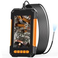
Endoscopes are medical devices used to visualize and examine the internal organs and structures of the body. They consist of a long, flexible tube with a light source and a camera at one end. The tube is inserted into the body through a natural opening or a small incision.
The light source illuminates the area being examined, while the camera captures real-time images or videos. These images are then transmitted to a monitor, allowing the healthcare professional to see the internal structures in detail. Some endoscopes also have channels that allow for the insertion of surgical instruments to perform procedures or collect samples.
Endoscopes can be used to examine various parts of the body, such as the gastrointestinal tract, respiratory system, urinary tract, and joints. They provide a minimally invasive way to diagnose and treat conditions, as they eliminate the need for large incisions and allow for direct visualization of the affected area.
1、 Optical Fiber Transmission
Endoscopes are medical devices used to visualize and examine the internal organs and cavities of the human body. They consist of a long, flexible tube with a light source and a camera at one end, which is inserted into the body through a natural opening or a small incision. The images captured by the camera are then transmitted to a monitor for real-time viewing by the medical professional.
One crucial component that enables the transmission of images in endoscopes is optical fiber technology. Optical fibers are thin, flexible strands made of high-quality glass or plastic that can transmit light over long distances with minimal loss of signal quality. These fibers are bundled together to form a fiber optic cable, which is integrated into the endoscope.
The light source at the end of the endoscope illuminates the area being examined, and the reflected light is collected by the camera. The camera converts the light signals into electrical signals, which are then transmitted through the optical fibers to the monitor. The monitor displays the captured images, allowing the medical professional to visualize and diagnose any abnormalities or conditions.
Optical fiber transmission in endoscopes offers several advantages. Firstly, it allows for high-resolution imaging, providing detailed and clear visuals of the internal organs. Secondly, the flexibility of the fibers enables the endoscope to navigate through the body's intricate pathways, reaching areas that would otherwise be difficult to access. Additionally, optical fibers are immune to electromagnetic interference, ensuring reliable and uninterrupted transmission of images.
In recent years, there have been advancements in endoscope technology, such as the development of smaller and more flexible fibers, allowing for even less invasive procedures. Furthermore, researchers are exploring the use of advanced imaging techniques, such as fluorescence and confocal microscopy, to enhance the diagnostic capabilities of endoscopes.
In conclusion, endoscopes rely on optical fiber transmission to capture and transmit high-quality images of the internal organs. This technology continues to evolve, enabling medical professionals to perform minimally invasive procedures and improve patient care.
2、 Illumination and Visualization
Endoscopes are medical devices used for visualizing and examining the internal organs and structures of the body. They consist of a long, flexible tube with a light source and a camera at one end, which allows doctors to see inside the body without the need for invasive surgery.
Illumination is a crucial aspect of endoscopes. The light source, typically a fiber optic cable or LED, is used to illuminate the area being examined. This allows the camera to capture clear and detailed images of the internal organs or structures. The light is transmitted through the endoscope's tube and directed towards the target area, providing visibility for the doctor.
Visualization is achieved through the camera attached to the endoscope. The camera captures real-time images or videos of the internal organs and sends them to a monitor, where the doctor can view and analyze them. The camera technology has evolved over the years, with the latest endoscopes utilizing high-definition cameras and advanced imaging techniques, such as narrow-band imaging or fluorescence imaging, to enhance visualization and improve diagnostic accuracy.
In recent years, there have been advancements in endoscope technology, such as the development of miniaturized endoscopes and capsule endoscopes. Miniaturized endoscopes are smaller in size and can be inserted through natural orifices, reducing patient discomfort and the need for sedation. Capsule endoscopes are swallowed by the patient and capture images as they pass through the digestive system, providing a non-invasive way to examine the small intestine.
Overall, endoscopes work by combining illumination and visualization to enable doctors to examine and diagnose various medical conditions without the need for invasive surgery. The continuous advancements in endoscope technology are improving patient care and expanding the range of applications for these devices.
3、 Flexible or Rigid Design
Flexible or Rigid Design
Endoscopes are medical devices used to visualize and examine the internal organs and structures of the body. They consist of a long, thin tube with a light source and a camera at one end, which allows doctors to see inside the body without the need for invasive surgery. There are two main types of endoscopes: flexible and rigid.
Flexible endoscopes are made up of a flexible tube that can be maneuvered through the body's natural passages, such as the digestive tract or respiratory system. The flexibility of these endoscopes allows for easier navigation through the curves and bends of the body, providing a more comprehensive examination. They are commonly used for procedures such as colonoscopies, bronchoscopies, and gastroscopies.
On the other hand, rigid endoscopes have a straight, inflexible tube and are typically used for procedures that require a more direct and precise approach, such as arthroscopies or laparoscopies. Rigid endoscopes provide a higher image quality due to the straight path of light transmission, but they require small incisions to access the targeted area.
In recent years, there have been advancements in endoscope technology, particularly in the field of flexible endoscopes. These advancements include the integration of high-definition cameras, improved lighting systems, and the development of miniaturized instruments that can be inserted through the endoscope to perform therapeutic procedures. Additionally, there has been progress in the development of wireless endoscopes, which eliminate the need for a physical connection between the endoscope and the imaging system.
Overall, endoscopes have revolutionized the field of medicine by providing a minimally invasive way to diagnose and treat various conditions. Whether flexible or rigid, these devices play a crucial role in improving patient outcomes and reducing the need for more invasive procedures.
4、 Camera and Image Capture
Endoscopes are medical devices used to visualize and examine the internal organs and structures of the body. They consist of a long, flexible tube with a light source and a camera at the tip. The camera captures images or videos of the internal body parts, allowing doctors to diagnose and treat various conditions.
The camera and image capture system in endoscopes play a crucial role in providing high-quality visuals. The camera is typically a small, high-resolution digital camera that can capture detailed images in real-time. The light source illuminates the area being examined, ensuring clear visibility for the camera.
The captured images or videos are then transmitted through the endoscope to a monitor, where the doctor can view them in real-time. This allows for immediate assessment and analysis of the patient's condition. In some cases, the images can also be recorded for future reference or shared with other medical professionals for consultation.
Advancements in technology have led to significant improvements in endoscope cameras and image capture systems. High-definition cameras with enhanced image sensors provide clearer and more detailed visuals. Some endoscopes even offer 3D imaging capabilities, allowing for a more immersive and accurate examination.
Furthermore, there have been recent developments in wireless endoscope systems, eliminating the need for physical cables and improving maneuverability during procedures. These wireless systems transmit the images or videos directly to a monitor or a mobile device, providing greater flexibility and convenience for doctors.
In conclusion, the camera and image capture system in endoscopes are essential components that enable doctors to visualize and examine internal body parts. Ongoing advancements in technology continue to enhance the quality and capabilities of endoscope cameras, improving diagnostic accuracy and patient care.


