How Do Light Microscopes Work A Level ?
Light microscopes work by using visible light to illuminate a specimen and magnify it for observation. The basic components of a light microscope include a light source, condenser, objective lens, eyepiece, and stage.
The light source, typically a bulb, produces light that passes through a condenser, which focuses and directs the light onto the specimen. The specimen is placed on the stage, which holds it in position for observation. The objective lens, located near the specimen, collects and magnifies the light that passes through the specimen. The magnified image is then further magnified by the eyepiece, which is where the observer looks through to view the specimen.
The magnification of a light microscope is determined by the combination of the objective lens and the eyepiece. The total magnification is calculated by multiplying the magnification of the objective lens by the magnification of the eyepiece. Additionally, the microscope may have various adjustments for focus, brightness, and contrast to optimize the image quality.
Overall, light microscopes allow for the visualization of small structures and details in a specimen by using visible light and a series of lenses to magnify the image.
1、 Principles of Light Microscopy
Light microscopes work based on the principles of optics and the interaction of light with the specimen being observed. These microscopes use visible light to illuminate the sample and magnify it for observation. The basic components of a light microscope include a light source, condenser, objective lens, eyepiece, and a stage to hold the specimen.
The light source, typically a halogen bulb, emits light that passes through a condenser. The condenser focuses and directs the light onto the specimen, which is placed on the stage. The objective lens, located near the specimen, collects the light that has interacted with the sample and forms an enlarged image. The eyepiece further magnifies this image, allowing the observer to view it.
The magnification power of a light microscope is determined by the combination of the objective lens and the eyepiece. The total magnification is calculated by multiplying the magnification of the objective lens by the magnification of the eyepiece. For example, if the objective lens has a magnification of 40x and the eyepiece has a magnification of 10x, the total magnification would be 400x.
In recent years, advancements in light microscopy have led to the development of techniques such as confocal microscopy and super-resolution microscopy. Confocal microscopy uses a pinhole to eliminate out-of-focus light, resulting in improved resolution and the ability to create three-dimensional images. Super-resolution microscopy techniques, such as stimulated emission depletion (STED) microscopy and structured illumination microscopy (SIM), surpass the diffraction limit of light, allowing for higher resolution imaging.
Overall, light microscopes work by utilizing the principles of optics to magnify and illuminate specimens, enabling scientists to observe and study a wide range of biological and non-biological samples.

2、 Components and Functionality of Light Microscopes
Light microscopes work by using visible light to magnify and resolve small objects or structures that are otherwise invisible to the naked eye. They consist of several key components that work together to produce a magnified image.
The first component is the light source, typically a bright white light or an LED, which provides illumination. The light passes through a condenser, which focuses and directs the light onto the specimen. The condenser also controls the amount of light that reaches the specimen, ensuring optimal contrast and resolution.
The specimen is placed on a glass slide and covered with a coverslip to protect it and provide a flat surface for viewing. The objective lens, located just below the stage, collects and magnifies the light that passes through the specimen. The magnification power of the objective lens can vary, typically ranging from 4x to 100x.
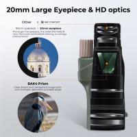
The magnified image formed by the objective lens is further magnified by the eyepiece or ocular lens, which is located at the top of the microscope. The eyepiece typically provides additional magnification, usually 10x, allowing the viewer to see a highly magnified image of the specimen.
Modern light microscopes often include additional features such as adjustable focus, fine focus controls, and various contrast-enhancing techniques like phase contrast or fluorescence microscopy. These advancements have greatly improved the resolution and clarity of images obtained using light microscopes.
In recent years, there have been significant advancements in light microscopy, such as the development of super-resolution techniques like stimulated emission depletion (STED) microscopy and structured illumination microscopy (SIM). These techniques allow for imaging at resolutions beyond the diffraction limit of light, enabling researchers to observe cellular structures and processes in unprecedented detail.
Overall, light microscopes function by using visible light and a combination of lenses to magnify and resolve small objects, providing valuable insights into the microscopic world.

3、 Illumination Techniques in Light Microscopy
Illumination techniques in light microscopy play a crucial role in the functioning of light microscopes. These techniques involve the manipulation of light to enhance the visibility and contrast of the specimen being observed.
One of the primary illumination techniques used in light microscopy is brightfield illumination. In this technique, a light source located beneath the specimen emits light, which passes through the condenser lens and is focused onto the specimen. The specimen absorbs some of the light, while the rest is transmitted through it. The objective lens then collects the transmitted light and forms an image that is magnified and projected onto the eyepiece for observation. Brightfield illumination is commonly used for observing stained or naturally pigmented specimens.
Another illumination technique is darkfield illumination, which is particularly useful for observing transparent or unstained specimens. In darkfield illumination, a special condenser lens is used to direct light at an oblique angle onto the specimen. This causes the light to scatter, making the specimen appear bright against a dark background. Darkfield illumination enhances the contrast and visibility of fine details in the specimen.
Phase contrast microscopy is another widely used illumination technique that allows for the observation of transparent specimens without the need for staining. This technique exploits the differences in refractive index within the specimen to create contrast. By using a special phase ring in the condenser and objective lenses, phase contrast microscopy converts the differences in refractive index into differences in brightness, making the specimen appear more distinct.
Fluorescence microscopy is a powerful illumination technique that utilizes fluorescent dyes or proteins to label specific structures within the specimen. These labels emit light of a different wavelength when excited by a specific wavelength of light. The emitted light is then collected by the objective lens and observed. Fluorescence microscopy allows for the visualization of specific molecules or structures within the specimen, providing valuable insights into cellular processes.
In recent years, advancements in light microscopy have led to the development of super-resolution techniques such as stimulated emission depletion (STED) microscopy and structured illumination microscopy (SIM). These techniques surpass the diffraction limit of light, enabling the observation of structures at a resolution previously thought to be unattainable with light microscopy.
In conclusion, illumination techniques in light microscopy are essential for enhancing the visibility and contrast of specimens. From traditional brightfield and darkfield illumination to more advanced techniques like phase contrast and fluorescence microscopy, these techniques have revolutionized our understanding of the microscopic world. The latest advancements in super-resolution microscopy have further expanded the capabilities of light microscopy, allowing for the visualization of structures at unprecedented levels of detail.

4、 Image Formation and Magnification in Light Microscopy
Light microscopes work by using visible light to illuminate a specimen and produce an enlarged image. The basic principle behind their operation is the interaction of light with the specimen, which allows for the formation of an image that can be observed by the human eye.
When light passes through the microscope, it first encounters the condenser, which focuses the light onto the specimen. The light then interacts with the specimen, either by being absorbed, transmitted, or reflected. The interaction of light with the specimen causes changes in its intensity, phase, or polarization, depending on the specific technique used.
The objective lens, located near the specimen, collects the light that has interacted with the specimen and forms a magnified real image. This image is further magnified by the eyepiece, which acts as a simple magnifying lens. The final image is observed by the human eye through the eyepiece.
The magnification of the image is determined by the combination of the objective lens and the eyepiece. The objective lens has a higher magnification power, typically ranging from 4x to 100x, while the eyepiece provides additional magnification, usually around 10x. The total magnification is calculated by multiplying the magnification of the objective lens by the magnification of the eyepiece.
In recent years, advancements in light microscopy have led to the development of techniques such as confocal microscopy, which allows for the acquisition of optical sections from thick specimens, and super-resolution microscopy, which surpasses the diffraction limit of light to achieve higher resolution. These techniques have revolutionized the field of microscopy, enabling researchers to observe biological structures and processes with unprecedented detail and clarity.


