How Does A Dissecting Microscope Work ?
A dissecting microscope, also known as a stereo microscope, works by using two separate optical paths to provide a three-dimensional view of the specimen being observed. The specimen is illuminated from above and below, and the light is reflected off the surface of the specimen and into the microscope's objective lenses. The lenses then magnify the image and project it into the eyepieces, where the observer can view the specimen in three dimensions. The two optical paths also provide a wider field of view than a traditional microscope, making it easier to observe larger specimens. The dissecting microscope is commonly used in biology, geology, and other fields where the observation of three-dimensional objects is important.
1、 Optical Design
How does a dissecting microscope work? The dissecting microscope, also known as a stereo microscope, is a type of microscope that is used to view objects in three dimensions. It works by using two separate optical paths to create a stereoscopic image of the specimen being viewed.
Optical Design: The dissecting microscope has two separate optical paths that are angled slightly differently. This allows the viewer to see the specimen from two slightly different angles, which creates a three-dimensional image. The two optical paths are created by two separate objective lenses, which are positioned at different angles to each other. The two images are then combined by the eyepiece, which allows the viewer to see the specimen in three dimensions.
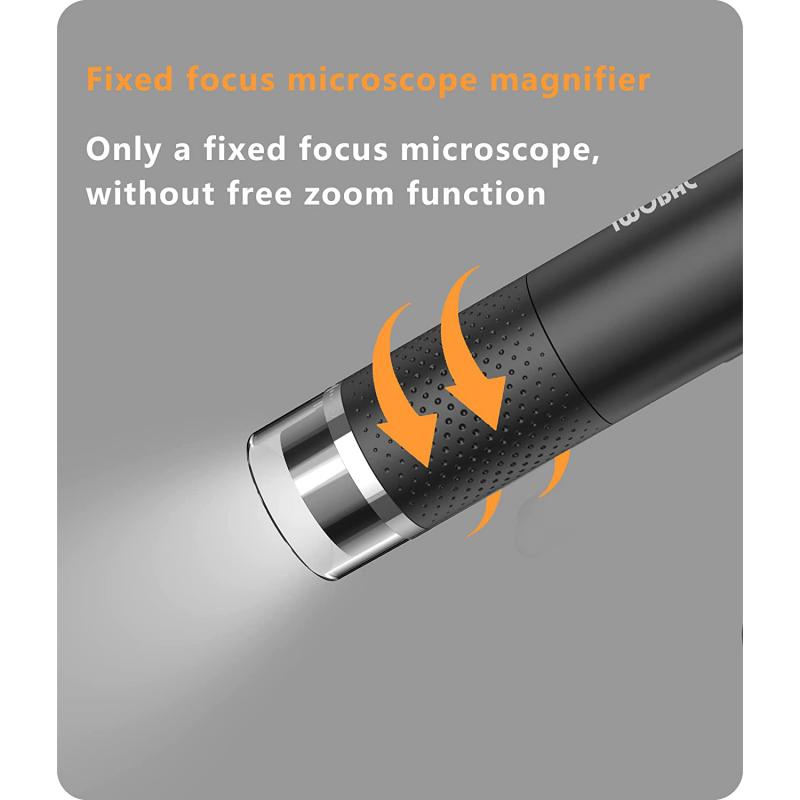
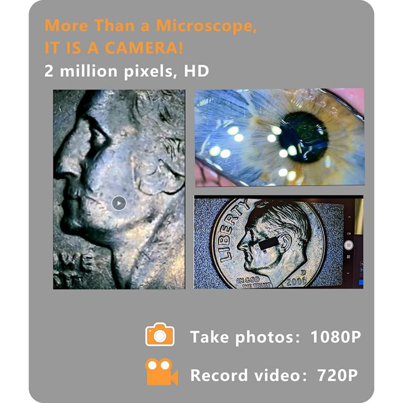
The latest point of view: In recent years, advances in technology have led to the development of digital dissecting microscopes. These microscopes use digital cameras to capture images of the specimen, which can then be viewed on a computer screen. This allows for greater magnification and more detailed images, as well as the ability to share images with others.
Overall, the dissecting microscope is an important tool for scientists and researchers in a variety of fields. Its ability to provide a three-dimensional view of specimens makes it particularly useful for studying complex structures and organisms. As technology continues to advance, we can expect to see even more sophisticated dissecting microscopes in the future.

2、 Magnification
A dissecting microscope, also known as a stereo microscope, is a type of microscope that is designed to provide a three-dimensional view of the specimen being observed. It works by using two separate optical paths that are angled slightly differently, which allows the viewer to see the specimen from two slightly different angles. This creates a three-dimensional image that is much easier to observe and study than a two-dimensional image.
One of the key features of a dissecting microscope is its magnification capabilities. These microscopes typically have a lower magnification range than other types of microscopes, such as compound microscopes. However, they make up for this by providing a much larger field of view, which allows the viewer to see more of the specimen at once.
In addition to its magnification capabilities, a dissecting microscope also has a number of other features that make it useful for a variety of applications. For example, many models have built-in lighting systems that provide illumination from above and below the specimen, which can help to highlight specific features or structures.
Overall, the dissecting microscope is an essential tool for anyone who needs to study specimens in three dimensions. Whether you are a biologist, a geologist, or a materials scientist, this type of microscope can help you to see your samples in a whole new way, and to gain insights that would be impossible to obtain with a traditional two-dimensional microscope.

3、 Illumination
How does a dissecting microscope work? Illumination is a key component of a dissecting microscope. The microscope uses a light source to illuminate the specimen being observed. The light is directed through the lenses of the microscope and onto the specimen, allowing the observer to see the specimen in greater detail.
The latest point of view on illumination in dissecting microscopes is the use of LED lights. LED lights are energy-efficient and have a longer lifespan than traditional light sources. They also produce less heat, which can be beneficial when observing live specimens.
In addition to illumination, a dissecting microscope also uses a system of lenses to magnify the specimen. The lenses are arranged in a way that allows the observer to view the specimen from different angles, providing a three-dimensional view.
The dissecting microscope is commonly used in biology, medicine, and other fields where the observation of small specimens is necessary. It is particularly useful for examining specimens that are too large or complex to be observed under a traditional microscope.
In conclusion, illumination is a crucial component of a dissecting microscope. The use of LED lights is the latest development in illumination technology for dissecting microscopes. The microscope's system of lenses and illumination work together to provide a detailed view of the specimen being observed.
4、 Focusing Mechanism
A dissecting microscope, also known as a stereo microscope, is a type of microscope that is used to view objects in three dimensions. It works by using two separate optical paths to create a stereoscopic image of the specimen being viewed. This allows the user to see the specimen in three dimensions, which is particularly useful for examining larger objects that cannot be viewed under a traditional compound microscope.
One of the key components of a dissecting microscope is the focusing mechanism. This mechanism allows the user to adjust the focus of the microscope to bring the specimen into sharp relief. There are several different types of focusing mechanisms that can be used in a dissecting microscope, including rack and pinion, coarse focus, and fine focus.
The rack and pinion mechanism is the most common type of focusing mechanism used in dissecting microscopes. It consists of a rack that is attached to the focus knob and a pinion gear that meshes with the rack. When the focus knob is turned, the rack moves up or down, which in turn moves the objective lens closer to or further away from the specimen.
The coarse focus mechanism is used to make large adjustments to the focus of the microscope. It typically consists of a large knob that is used to move the objective lens up or down. The fine focus mechanism, on the other hand, is used to make small adjustments to the focus of the microscope. It typically consists of a smaller knob that is used to move the objective lens in very small increments.
In recent years, advances in technology have led to the development of digital dissecting microscopes. These microscopes use digital cameras and software to create three-dimensional images of the specimen being viewed. This allows for even greater precision and accuracy in examining specimens, and has led to new discoveries in a variety of fields, including biology, medicine, and materials science.
