How Does A Light Microscope Work ?
A light microscope works by using visible light to magnify and resolve small objects or structures that are otherwise invisible to the naked eye. The light passes through a series of lenses, including an objective lens and an eyepiece lens, which work together to magnify the image of the specimen. The objective lens is positioned close to the specimen and collects the light that is reflected or transmitted through it. The light is then focused onto the eyepiece lens, which further magnifies the image and projects it onto the observer's eye or a camera.
The resolution of a light microscope is limited by the wavelength of visible light, which is around 400-700 nanometers. This means that it can only resolve structures that are larger than this size. To improve the resolution, various techniques such as staining, phase contrast, and fluorescence microscopy can be used. Light microscopes are commonly used in biology, medicine, and materials science to study cells, tissues, and materials at the microscale level.
1、 Optical components
How does a light microscope work? Light microscopes use a combination of optical components to magnify and focus light onto a specimen. The basic components of a light microscope include the objective lens, eyepiece, stage, and light source. The objective lens is the primary lens that magnifies the specimen, while the eyepiece further magnifies the image for the viewer. The stage holds the specimen in place and allows for precise movement, while the light source illuminates the specimen.
When light passes through the objective lens, it is refracted and focused onto the specimen. The light then passes through the specimen and is refracted again as it passes through the objective lens. This creates an enlarged and inverted image of the specimen, which is then magnified further by the eyepiece.
Recent advancements in light microscopy have allowed for even higher resolution and more detailed imaging. Techniques such as confocal microscopy and super-resolution microscopy use specialized optical components and computer algorithms to produce images with greater clarity and detail. These techniques have revolutionized the field of biology and have allowed researchers to study cellular structures and processes in unprecedented detail.
In summary, light microscopes work by using a combination of optical components to magnify and focus light onto a specimen. Recent advancements in microscopy have allowed for even higher resolution and more detailed imaging, leading to new discoveries and insights in the field of biology.

2、 Magnification and resolution
A light microscope works by using visible light to magnify and resolve small objects or structures that are otherwise invisible to the naked eye. The light passes through a series of lenses, which bend and focus the light to create a magnified image of the specimen. The magnification is determined by the combination of the objective lens and the eyepiece lens, which work together to enlarge the image.
Magnification is the process of making an object appear larger than its actual size. The light microscope can magnify objects up to 1000 times their original size, allowing scientists to study the fine details of cells, tissues, and other small structures. However, magnification alone is not enough to provide a clear image of the specimen.
Resolution is the ability to distinguish between two closely spaced objects. The light microscope's resolution is limited by the wavelength of visible light, which is about 500 nanometers. This means that two objects closer than 500 nanometers will appear as a single blurred image. To improve resolution, scientists use techniques such as staining, which highlights specific structures within the specimen, and confocal microscopy, which uses a laser to scan the specimen and create a 3D image.
In recent years, advancements in technology have led to the development of super-resolution microscopy, which can achieve resolutions beyond the diffraction limit of visible light. This technique uses fluorescent molecules to create a highly detailed image of the specimen, allowing scientists to study the structure and function of cells and tissues at the molecular level.
In conclusion, the light microscope works by using visible light to magnify and resolve small objects. Magnification and resolution are the two key factors that determine the quality of the image produced by the microscope. With the latest advancements in technology, scientists can now study the intricate details of cells and tissues at the molecular level, opening up new avenues for research and discovery.

3、 Illumination sources
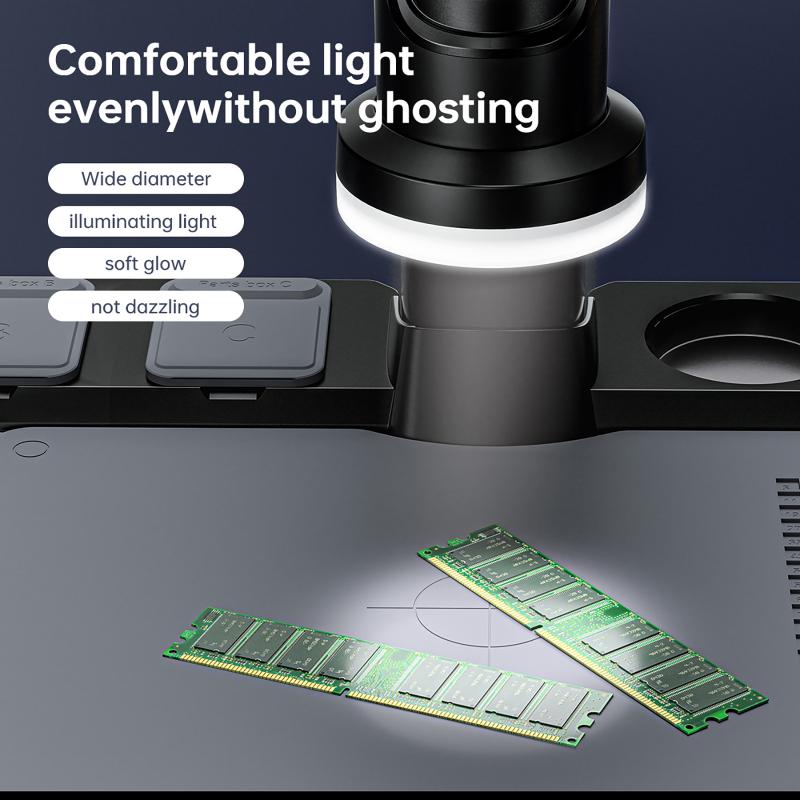
How does a light microscope work? One of the key components of a light microscope is the illumination source. This source provides the light that is used to illuminate the sample being observed. In most modern light microscopes, the illumination source is typically a high-intensity LED or halogen bulb.
The light from the illumination source passes through a series of lenses and filters before reaching the sample. These lenses and filters help to focus and shape the light, as well as remove any unwanted wavelengths or reflections.
Once the light reaches the sample, it interacts with the various structures and molecules within the sample. Some of the light is absorbed, while other light is scattered or refracted. This interaction between the light and the sample is what allows us to see the sample under the microscope.
The light that passes through the sample then enters the objective lens, which is the lens closest to the sample. The objective lens magnifies the image of the sample and projects it onto the eyepiece or camera.
In recent years, there have been significant advancements in the field of light microscopy. For example, the development of super-resolution microscopy techniques has allowed researchers to visualize structures and molecules at a resolution that was previously thought to be impossible with light microscopy. Additionally, the use of fluorescent probes and dyes has enabled researchers to selectively label specific structures or molecules within a sample, further enhancing the capabilities of light microscopy.

4、 Sample preparation
How does a light microscope work?
A light microscope works by using visible light to magnify and resolve small objects. The light passes through the specimen and is refracted by the lenses in the microscope, which magnify the image and project it onto the eyepiece or camera. The quality of the image depends on the quality of the lenses and the amount of light that is able to pass through the specimen.
Sample preparation is an important step in using a light microscope. The specimen must be thin enough to allow light to pass through it, and it must be mounted on a slide in a way that preserves its structure and prevents it from drying out or being damaged. Depending on the type of specimen, different techniques may be used to prepare it for observation, such as staining, sectioning, or fixing.
Recent advances in light microscopy have allowed for even higher resolution and more detailed imaging of biological specimens. Techniques such as confocal microscopy, super-resolution microscopy, and live-cell imaging have revolutionized the field of cell biology and allowed researchers to study cellular processes in unprecedented detail. These techniques have also been combined with other imaging modalities, such as electron microscopy and X-ray crystallography, to provide a more complete understanding of the structure and function of biological molecules and systems.


