How Does A Medical Endoscope Work ?
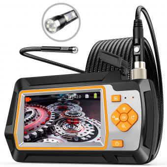
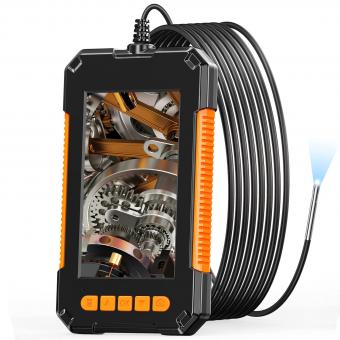
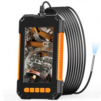
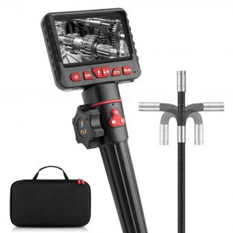
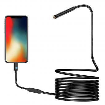
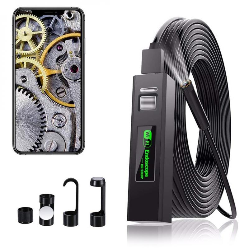
A medical endoscope is a flexible tube with a light and a camera at its tip. It is inserted into the body through a natural opening or a small incision. The light illuminates the area being examined, while the camera captures images and sends them to a monitor for visualization. The endoscope can be maneuvered to navigate through the body's internal structures, allowing doctors to visually inspect organs, tissues, or cavities. Some endoscopes also have channels that enable the insertion of surgical instruments for procedures such as biopsies or removal of abnormal tissue. Overall, the medical endoscope provides a minimally invasive way to diagnose and treat various medical conditions.
1、 Optical Fiber Transmission
A medical endoscope is a vital tool used in modern medicine for diagnosing and treating various medical conditions. It allows doctors to visualize and examine internal organs and structures without the need for invasive surgery. One of the key components that enables the functionality of a medical endoscope is optical fiber transmission.
Optical fiber transmission involves the use of thin, flexible glass or plastic fibers that can transmit light signals over long distances with minimal loss of intensity. In the case of a medical endoscope, these fibers are bundled together and integrated into the device. The light source, typically a high-intensity LED or laser, is connected to one end of the fiber bundle, while the other end is attached to a camera or eyepiece for visualization.
When the light source is activated, it emits a beam of light that travels through the fiber bundle. The light is then transmitted through the endoscope's insertion tube, which is inserted into the patient's body. The light illuminates the internal structures, and the reflected light is captured by the camera or eyepiece, allowing the doctor to see real-time images of the area being examined.
Optical fiber transmission offers several advantages in medical endoscopy. Firstly, it allows for the transmission of light over long distances, enabling the endoscope to reach deep into the body. Secondly, the flexibility of the fibers allows for easy maneuverability within the body, minimizing patient discomfort. Additionally, the use of optical fibers ensures that the light signal remains clear and undistorted, providing high-quality images for accurate diagnosis.
In recent years, there have been advancements in endoscopic technology, such as the development of smaller and more flexible fibers, as well as the integration of advanced imaging techniques like fluorescence and confocal microscopy. These advancements have further improved the capabilities of medical endoscopes, allowing for better visualization and detection of abnormalities.
In conclusion, optical fiber transmission is a crucial aspect of how a medical endoscope works. It enables the transmission of light signals, providing illumination and visualization of internal structures. With ongoing advancements in endoscopic technology, medical professionals can continue to benefit from improved imaging capabilities, leading to more accurate diagnoses and better patient outcomes.
2、 Illumination System
The Illumination System in a medical endoscope plays a crucial role in providing clear and well-lit images of the internal body structures. It consists of various components that work together to ensure optimal illumination during endoscopic procedures.
The primary component of the Illumination System is the light source, which can be either a halogen or xenon lamp. These lamps emit intense light that is transmitted through a fiber optic cable to the distal end of the endoscope. At the distal end, the light is evenly distributed by a diffuser or a lens system to illuminate the area of interest.
To achieve the best possible illumination, the light is often filtered to remove any unwanted wavelengths or colors. Filters can be used to enhance contrast or to selectively highlight certain tissues or structures. Additionally, the intensity of the light can be adjusted to suit the specific requirements of the procedure.
In recent years, there have been advancements in endoscope illumination technology. LED (Light Emitting Diode) light sources are increasingly being used due to their compact size, energy efficiency, and longer lifespan compared to traditional lamps. LED-based illumination systems offer improved color rendering and can be easily integrated into portable and wireless endoscope devices.
Furthermore, some endoscopes now incorporate advanced imaging technologies such as narrow-band imaging (NBI) or fluorescence imaging. NBI utilizes specific light wavelengths to enhance the visualization of blood vessels and surface structures, while fluorescence imaging involves the use of fluorescent dyes to highlight abnormal tissues or lesions.
In conclusion, the Illumination System in a medical endoscope is responsible for providing adequate and well-controlled light to visualize internal body structures during endoscopic procedures. Ongoing advancements in light sources and imaging technologies continue to improve the quality and accuracy of endoscopic imaging, enabling better diagnosis and treatment outcomes.
3、 Camera and Image Processing
A medical endoscope is a device used by healthcare professionals to visualize and examine the internal organs and structures of the body. It consists of a long, flexible tube with a light source and a camera at one end. The other end of the endoscope is inserted into the body through a natural opening or a small incision.
The camera and image processing play a crucial role in the functioning of a medical endoscope. The camera captures high-resolution images or videos of the internal structures, which are then processed and displayed on a monitor for the healthcare professional to analyze.
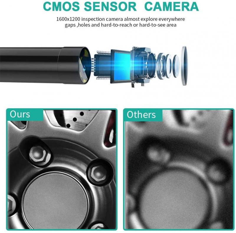
The camera in a medical endoscope is typically a charge-coupled device (CCD) or a complementary metal-oxide-semiconductor (CMOS) sensor. These sensors convert the light signals received from the internal structures into electrical signals. The electrical signals are then processed by the image processing unit of the endoscope.
The image processing unit enhances the captured images by adjusting the brightness, contrast, and color levels. It also removes any noise or artifacts to provide a clear and detailed view of the internal structures. The processed images are then transmitted to a monitor in real-time, allowing the healthcare professional to observe and diagnose any abnormalities or diseases.
In recent years, there have been advancements in endoscopic technology, such as the development of high-definition (HD) and ultra-high-definition (4K) cameras. These cameras provide even greater clarity and detail, enabling healthcare professionals to detect and diagnose conditions with higher accuracy.
Furthermore, there have been advancements in image processing algorithms, including artificial intelligence (AI) and machine learning techniques. These algorithms can analyze the captured images and assist healthcare professionals in detecting and classifying abnormalities, potentially improving diagnostic accuracy and efficiency.
Overall, the camera and image processing components of a medical endoscope are essential for visualizing and analyzing the internal structures of the body, aiding in the diagnosis and treatment of various medical conditions.

4、 Flexible or Rigid Shaft
A medical endoscope is a device used by healthcare professionals to visualize and examine the internal organs and structures of the body. It consists of a long, thin tube with a light source and a camera at one end, which allows for real-time imaging of the area being examined. The other end of the endoscope is connected to a monitor, where the images are displayed for the healthcare provider to analyze.
There are two main types of medical endoscopes: flexible and rigid shaft. The choice between the two depends on the specific procedure and the area of the body being examined.
Flexible endoscopes are made of a flexible fiber-optic bundle that allows for easy maneuverability through the body's natural curves and bends. They are commonly used for procedures such as colonoscopy, upper gastrointestinal endoscopy, and bronchoscopy. The flexibility of these endoscopes enables them to reach areas that may be difficult to access with a rigid endoscope.
On the other hand, rigid shaft endoscopes are more commonly used for procedures that require a straight path, such as arthroscopy (joint examination) or laparoscopy (abdominal examination). These endoscopes have a rigid tube with lenses and a light source, providing high-quality images.
In recent years, there have been advancements in endoscope technology. One notable development is the introduction of miniaturized endoscopes, which are smaller in size and can be used for procedures in narrow or delicate areas of the body. Additionally, there have been improvements in image quality, with high-definition cameras being incorporated into endoscopes, allowing for clearer and more detailed visualization.
Overall, medical endoscopes play a crucial role in diagnosing and treating various medical conditions. They provide healthcare professionals with a non-invasive means of examining internal structures, aiding in accurate diagnoses and guiding minimally invasive procedures.
