How Does A Microscope Work ?
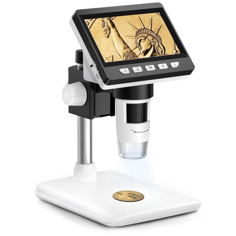
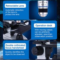
A microscope works by using lenses to magnify small objects or organisms that are not visible to the naked eye. Light microscopes, the most common type, use a combination of lenses to focus light onto the specimen. The objective lens collects and magnifies the light, while the eyepiece lens further magnifies the image for the viewer. The specimen is placed on a glass slide and illuminated from below with a light source. As light passes through the specimen, it interacts with the structures or cells, causing them to scatter or absorb the light. This creates contrast and allows the viewer to see the specimen more clearly. The magnified image is then observed through the eyepiece and can be further adjusted using focus knobs to bring the specimen into sharp focus. More advanced microscopes, such as electron microscopes, use beams of electrons instead of light to achieve even higher magnification and resolution.
1、 Optical Microscopy: Utilizes lenses to magnify and focus light.
Optical microscopy is a widely used technique that allows scientists and researchers to observe and study objects that are too small to be seen with the naked eye. It works by utilizing lenses to magnify and focus light, enabling the visualization of tiny details and structures.
The basic principle behind optical microscopy involves the interaction of light with the specimen being observed. When light passes through the lens system of a microscope, it undergoes refraction, which causes the light rays to converge and form an enlarged image of the specimen. This image is then further magnified by additional lenses, such as the eyepiece, allowing the observer to see the object in greater detail.
In a traditional light microscope, a light source, such as a bulb, illuminates the specimen from below, while the lenses in the microscope system focus the light onto the sample. The light interacts with the specimen, either by being absorbed, transmitted, or reflected, depending on the properties of the object. The resulting image is then magnified and projected into the observer's eye, either directly or through a camera.
Recent advancements in optical microscopy have led to the development of various techniques that enhance the resolution and capabilities of traditional microscopes. For example, confocal microscopy uses a pinhole aperture to eliminate out-of-focus light, resulting in sharper images and improved optical sectioning. Super-resolution microscopy techniques, such as stimulated emission depletion (STED) microscopy and structured illumination microscopy (SIM), have pushed the limits of resolution beyond the diffraction limit, allowing for the visualization of even smaller structures.
In summary, optical microscopy works by utilizing lenses to magnify and focus light, enabling the observation of objects that are too small to be seen with the naked eye. Ongoing advancements in microscopy techniques continue to expand our understanding of the microscopic world and drive scientific discoveries in various fields.

2、 Electron Microscopy: Uses electron beams to visualize ultra-small structures.
A microscope is an instrument used to magnify and visualize objects that are too small to be seen with the naked eye. It works by using a combination of lenses or electron beams to focus light or electrons onto the object being observed.
In the case of a light microscope, it uses a series of lenses to magnify the image of the object. The light source at the base of the microscope illuminates the object, and the lenses in the eyepiece and objective lens system work together to magnify and focus the image onto the retina of the observer's eye. This allows for the visualization of objects at a much higher magnification than what is possible with the human eye alone.
On the other hand, electron microscopy takes the concept of microscopy to a whole new level. Instead of using light, it uses a beam of electrons to visualize ultra-small structures. Electron microscopes have a much higher resolution than light microscopes, allowing for the observation of objects at the atomic level. The electron beam is focused onto the object using electromagnetic lenses, and the resulting image is captured on a fluorescent screen or a digital detector.
The latest advancements in electron microscopy include techniques such as scanning electron microscopy (SEM) and transmission electron microscopy (TEM). SEM provides detailed surface information by scanning the object with a focused electron beam and detecting the emitted secondary electrons. TEM, on the other hand, allows for the visualization of the internal structure of the object by transmitting the electron beam through a thin section of the sample.
In summary, microscopes work by using lenses or electron beams to magnify and focus light or electrons onto the object being observed. The latest advancements in electron microscopy have revolutionized our ability to visualize and understand ultra-small structures.

3、 Confocal Microscopy: Captures images by scanning laser beams through a pinhole.
A microscope is an essential tool used in scientific research and medical diagnostics to observe objects that are too small to be seen with the naked eye. It works by utilizing the principles of optics to magnify and enhance the visibility of tiny structures.
One type of microscope that has gained significant popularity in recent years is the confocal microscope. Confocal microscopy captures images by scanning laser beams through a pinhole. This technique allows for the precise focusing of light onto a specific plane within a sample, resulting in improved resolution and contrast.
In a confocal microscope, the sample is illuminated with a laser beam, which is then focused onto a specific point within the sample. The light that is reflected or emitted from the sample is collected by a detector, and a computer generates an image based on the intensity of the detected light. By scanning the laser beam across the sample, a series of images can be obtained, which can then be reconstructed into a three-dimensional representation of the sample.
The advantage of confocal microscopy lies in its ability to eliminate out-of-focus light, resulting in sharper images with improved clarity and contrast. This technique has revolutionized the field of biological imaging, allowing researchers to study intricate cellular structures and dynamic processes in unprecedented detail.
In recent years, advancements in confocal microscopy have further enhanced its capabilities. For example, the development of super-resolution techniques, such as stimulated emission depletion (STED) microscopy and structured illumination microscopy (SIM), has pushed the limits of resolution beyond the diffraction limit of light. These techniques enable researchers to visualize structures at the nanoscale level, opening up new avenues for understanding biological processes.
Overall, the use of confocal microscopy and its continuous advancements have greatly contributed to our understanding of the microscopic world and have paved the way for groundbreaking discoveries in various scientific disciplines.

4、 Scanning Probe Microscopy: Measures surface properties using a physical probe.
Scanning Probe Microscopy (SPM) is a powerful technique used to measure surface properties at the nanoscale level. Unlike traditional optical microscopes that rely on light to magnify and visualize samples, SPM utilizes a physical probe to interact with the surface of the sample.
The basic principle behind SPM is the scanning of a sharp probe over the sample surface while measuring various properties. The probe, typically a sharp tip made of materials like silicon or diamond, is attached to a cantilever. The cantilever acts as a spring, allowing the probe to move up and down as it interacts with the surface.
There are several types of SPM techniques, including Atomic Force Microscopy (AFM) and Scanning Tunneling Microscopy (STM). AFM measures the forces between the probe and the sample surface, while STM measures the tunneling current between the probe and the sample.
In AFM, as the probe scans the surface, it experiences attractive or repulsive forces between the atoms on the probe and the atoms on the sample surface. These forces cause the cantilever to deflect, and this deflection is measured using a laser beam reflected off the cantilever. By mapping the deflection, a three-dimensional image of the surface can be generated.
In STM, a small bias voltage is applied between the probe and the sample, creating a tunneling current. The current is highly sensitive to the distance between the probe and the sample surface. By scanning the probe over the surface while maintaining a constant current, a topographic image of the surface can be obtained.
The latest advancements in SPM techniques have allowed for even higher resolution imaging and the ability to measure various properties such as electrical conductivity, magnetic fields, and chemical composition. Additionally, new techniques like non-contact AFM and dynamic AFM have been developed to minimize the interaction forces between the probe and the sample, enabling imaging of delicate samples and even single molecules.
Overall, SPM provides a powerful tool for studying and manipulating materials at the nanoscale, allowing researchers to explore the world of atoms and molecules with unprecedented detail and precision.


