How Does A Raman Microscope Work ?
A Raman microscope works by utilizing the Raman scattering phenomenon. It involves the interaction of light with a sample, where a small fraction of the incident light undergoes inelastic scattering. This scattering results in a shift in the energy of the scattered photons, which corresponds to the vibrational energy levels of the sample molecules.
In a Raman microscope, a laser beam is focused onto the sample, and the scattered light is collected and analyzed. The collected light is passed through a spectrometer, which separates the different wavelengths of light. The spectrometer then detects and measures the intensity of the scattered light at different wavelengths.
By analyzing the Raman scattered light, the microscope can provide information about the molecular composition, structure, and vibrational modes of the sample. This allows for the identification and characterization of various materials, including solids, liquids, and gases, at a microscopic level. Raman microscopy finds applications in various fields, such as materials science, chemistry, biology, and forensics.
1、 Raman Scattering: Inelastic scattering of light to analyze molecular vibrations.
A Raman microscope is an analytical tool that utilizes Raman scattering to analyze molecular vibrations. Raman scattering is an inelastic scattering of light, where the incident photons interact with the sample molecules and undergo a change in energy. This change in energy corresponds to the vibrational modes of the molecules, providing valuable information about their chemical composition and structure.
In a Raman microscope, a laser beam is focused onto the sample, and the scattered light is collected and analyzed. The majority of the scattered light is elastically scattered, meaning it has the same energy as the incident laser beam. However, a small fraction of the scattered light undergoes Raman scattering, resulting in a shift in energy. This energy shift is measured and used to determine the vibrational modes of the molecules.
The Raman microscope typically employs a spectrometer to analyze the scattered light. The spectrometer separates the different wavelengths of light, allowing the detection of the Raman-shifted photons. By comparing the energy shift of the scattered light with a known reference, the vibrational modes of the sample molecules can be identified.
The latest advancements in Raman microscopy have focused on improving the sensitivity and spatial resolution of the technique. This has been achieved through the use of advanced laser sources, such as near-infrared lasers, which can enhance the Raman signal and reduce fluorescence background. Additionally, the integration of confocal microscopy techniques allows for precise spatial localization of the Raman signal, enabling the analysis of specific regions within a sample.
Overall, a Raman microscope provides a non-destructive and label-free method for analyzing molecular vibrations, making it a powerful tool in various fields, including materials science, pharmaceuticals, and biological research.
2、 Laser Excitation: Use of a laser to excite the sample.
A Raman microscope is an advanced analytical tool that utilizes the Raman scattering phenomenon to provide detailed information about the molecular composition and structure of a sample. Raman spectroscopy is a non-destructive technique that can be used to analyze a wide range of materials, including solids, liquids, and gases.
The basic principle behind a Raman microscope is the interaction between a laser beam and the sample. When a laser beam is directed onto a sample, a small fraction of the incident photons undergoes inelastic scattering, resulting in a shift in energy known as the Raman effect. This shift in energy corresponds to the vibrational modes of the molecules present in the sample.
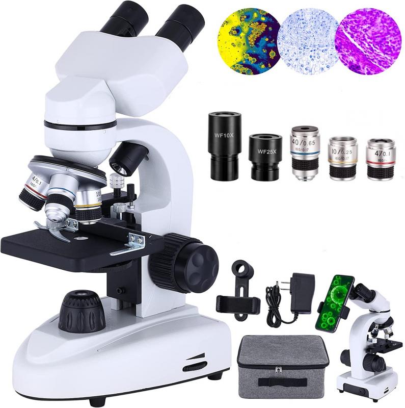
The Raman microscope consists of several key components, including a laser source, a microscope objective, a spectrometer, and a detector. Laser Excitation is the first step in the process, where a laser beam is focused onto the sample. The laser excitation provides the energy required to induce the Raman scattering.
The scattered light from the sample is collected by the microscope objective and directed towards the spectrometer. The spectrometer separates the scattered light into its different wavelengths using a diffraction grating or a prism. The dispersed light is then detected by a sensitive detector, such as a charge-coupled device (CCD) or a photomultiplier tube (PMT).
By analyzing the Raman scattered light, the Raman microscope can provide valuable information about the molecular composition, chemical bonding, and structural characteristics of the sample. This information can be used in various fields, including materials science, pharmaceuticals, forensics, and environmental analysis.
In recent years, advancements in technology have led to the development of more compact and portable Raman microscopes. These instruments offer improved sensitivity, faster data acquisition, and enhanced spatial resolution. Additionally, the integration of other imaging techniques, such as fluorescence microscopy, has allowed for multimodal imaging, providing a more comprehensive understanding of the sample.
Overall, a Raman microscope works by utilizing laser excitation to induce Raman scattering, which is then analyzed to obtain valuable molecular information about the sample. The continuous advancements in Raman microscopy technology are expanding its applications and making it an indispensable tool in various scientific and industrial fields.
3、 Spectral Analysis: Measurement and analysis of the scattered light spectrum.
A Raman microscope is an advanced analytical tool that utilizes the Raman scattering phenomenon to provide detailed information about the molecular composition and structure of a sample. Raman spectroscopy is a non-destructive technique that can be used to analyze a wide range of materials, including solids, liquids, and gases.
The working principle of a Raman microscope involves the measurement and analysis of the scattered light spectrum. When a sample is illuminated with a laser beam, a small fraction of the incident light is scattered inelastically due to the interaction with the sample's molecules. This scattered light contains information about the vibrational and rotational energy levels of the molecules, which can be used to identify the chemical composition and molecular structure of the sample.
In a Raman microscope, the scattered light is collected and focused onto a spectrometer. The spectrometer separates the different wavelengths of light and measures their intensities. By analyzing the spectrum of the scattered light, it is possible to identify the specific molecular vibrations and obtain a fingerprint of the sample.
The latest advancements in Raman microscopy include the integration of high-resolution imaging capabilities. This allows for the visualization of the spatial distribution of different molecular species within a sample. Additionally, techniques such as confocal Raman microscopy and surface-enhanced Raman spectroscopy (SERS) have been developed to enhance the sensitivity and spatial resolution of Raman measurements.
Overall, a Raman microscope provides a powerful tool for chemical analysis and imaging, offering valuable insights into the molecular composition and structure of a wide range of materials.
4、 Molecular Identification: Identification of molecules based on their unique Raman spectra.
A Raman microscope is an advanced analytical tool used for molecular identification based on the unique Raman spectra of molecules. Raman spectroscopy, discovered by Sir C.V. Raman in 1928, involves the interaction of light with matter, resulting in the scattering of photons at different wavelengths. This scattering phenomenon provides valuable information about the molecular composition and structure of a sample.
In a Raman microscope, a laser beam is focused onto the sample, causing the molecules to vibrate and scatter light at different frequencies. The scattered light is collected and analyzed using a spectrometer, which separates the different wavelengths of light. The resulting Raman spectrum is a unique fingerprint of the sample, providing information about the molecular bonds, functional groups, and overall chemical composition.
The Raman microscope offers several advantages over other analytical techniques. It is non-destructive, requiring minimal sample preparation, and can be used to analyze a wide range of materials, including solids, liquids, and gases. Additionally, it provides high spatial resolution, allowing for the identification of molecules in specific regions of a sample.
Recent advancements in Raman microscopy have further enhanced its capabilities. For example, the integration of confocal microscopy allows for three-dimensional imaging and mapping of molecular distributions within a sample. Additionally, the use of advanced data analysis techniques, such as machine learning algorithms, has improved the speed and accuracy of molecular identification.
In summary, a Raman microscope works by utilizing the unique Raman spectra of molecules to identify and analyze samples. Its non-destructive nature, versatility, and high spatial resolution make it a powerful tool in various fields, including materials science, pharmaceuticals, forensics, and biology. Ongoing advancements continue to expand the capabilities of Raman microscopy, making it an invaluable tool for molecular identification and characterization.
