How Does An Endoscope Work ?
An endoscope is a medical device used to visualize and examine the internal organs and structures of the body. It consists of a long, flexible tube with a light source and a camera at one end. The tube is inserted into the body through a natural opening or a small incision.
The light source illuminates the area being examined, allowing the camera to capture high-quality images or videos. These images are then transmitted to a monitor, where the healthcare professional can view them in real-time. Some endoscopes also have channels that allow for the insertion of surgical instruments to perform procedures or collect tissue samples.
The flexibility of the endoscope allows it to navigate through the body's curves and bends, reaching areas that would otherwise be difficult to access. The camera provides a detailed view of the internal structures, helping in the diagnosis and treatment of various medical conditions. Endoscopes are commonly used in procedures such as gastrointestinal examinations, bronchoscopy, arthroscopy, and laparoscopy.
1、 Optical Fiber Transmission
An endoscope is a medical device used to visualize and examine the internal organs and cavities of the human body. It consists of a long, flexible tube with a light source and a camera at one end, and a viewing screen at the other end. The endoscope is inserted into the body through a natural opening or a small incision, allowing doctors to see and diagnose various conditions.
One of the key components that enables the functioning of an endoscope is optical fiber transmission. Optical fibers are thin, flexible strands made of high-quality glass or plastic that can transmit light over long distances with minimal loss. In the case of an endoscope, optical fibers are used to transmit light from the light source to illuminate the area being examined.
The light source, typically a bright LED or a halogen lamp, emits light that is guided through the optical fibers. The light is then directed towards the tip of the endoscope, where it is emitted to illuminate the internal organs or cavities. The reflected light from the illuminated area is captured by the camera at the end of the endoscope and transmitted back through the optical fibers to the viewing screen.
The viewing screen displays the real-time images captured by the camera, allowing doctors to visualize and examine the internal structures. The images can also be recorded for further analysis or documentation.
Optical fiber transmission in endoscopes offers several advantages. It allows for flexible and maneuverable endoscopes, as the thin optical fibers can be easily bent and routed through the body. It also provides high-quality, high-resolution images, enabling accurate diagnosis and treatment. Additionally, optical fibers are immune to electromagnetic interference, ensuring reliable transmission of light signals.
In recent years, there have been advancements in endoscope technology, such as the development of smaller and more flexible endoscopes, as well as the integration of advanced imaging techniques like fluorescence and confocal microscopy. These advancements aim to improve the diagnostic capabilities of endoscopes and enhance patient care.
2、 Illumination System
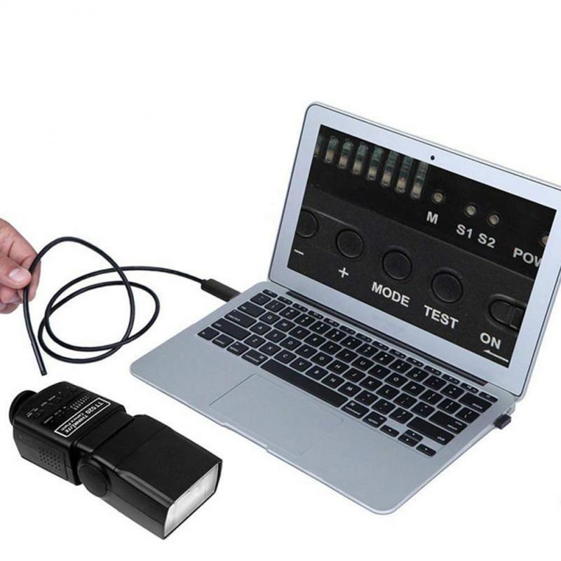
The Illumination System in an endoscope plays a crucial role in providing clear and well-lit images during medical procedures. It consists of various components that work together to ensure optimal illumination of the target area.
The primary component of the Illumination System is the light source, which can be either a halogen or xenon lamp. These lamps emit intense light that is then transmitted through a fiber optic cable. The cable carries the light to the distal end of the endoscope, where it is dispersed evenly across the target area.
At the distal end, the light is directed towards the target tissue through a series of lenses and mirrors. These optical elements help focus and shape the light beam, ensuring that it illuminates the desired area effectively. Additionally, some endoscopes may also have adjustable light intensity settings to provide flexibility in different clinical scenarios.
In recent years, there have been advancements in endoscope illumination technology. One notable development is the integration of light-emitting diodes (LEDs) as the light source. LEDs offer several advantages over traditional lamps, including longer lifespan, lower power consumption, and a wider range of color temperatures. LED-based illumination systems also allow for better control of light intensity and color, enabling enhanced visualization of tissues and structures.
Furthermore, the use of advanced imaging sensors and image processing algorithms has improved the overall image quality in endoscopy. These technologies work in conjunction with the Illumination System to capture and enhance the visual information, providing clinicians with clearer and more detailed images for accurate diagnosis and treatment.
In conclusion, the Illumination System in an endoscope is a critical component that ensures proper lighting of the target area. With advancements in LED technology and image processing, endoscopes now offer improved illumination and image quality, enabling healthcare professionals to perform procedures with greater precision and accuracy.
3、 Lens System
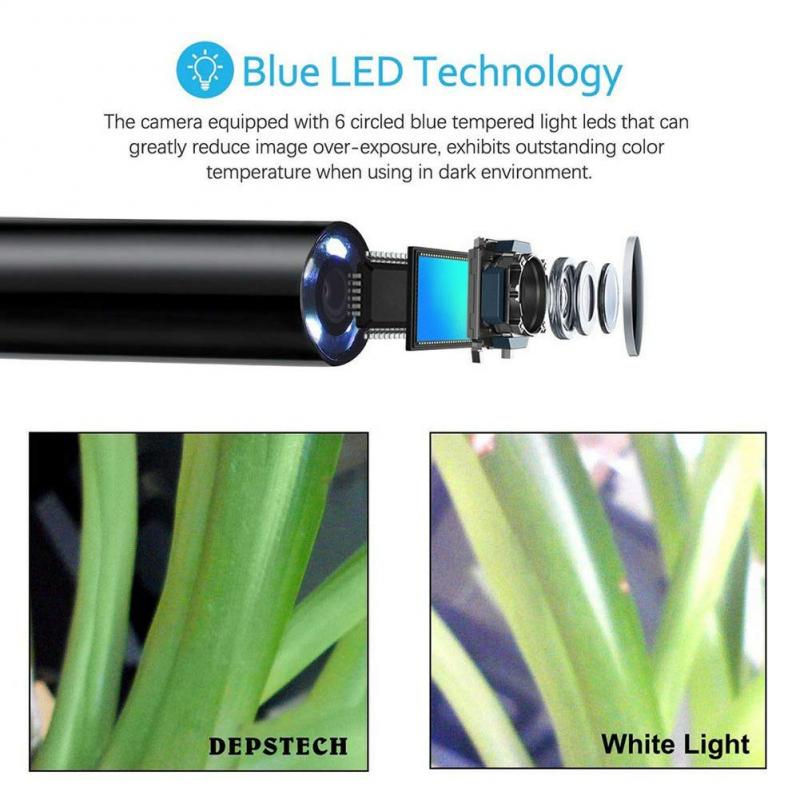
An endoscope is a medical device used to visualize and examine the internal organs and structures of the body. It consists of a long, flexible tube with a light source and a camera at one end, and a lens system at the other end. The lens system is a crucial component of the endoscope as it allows for the magnification and focusing of the image.
The lens system in an endoscope works similarly to the lens system in a camera. It is composed of multiple lenses that are arranged in a specific configuration to capture and transmit light. When light enters the endoscope through the patient's body, it passes through the lenses, which focus the light onto the camera sensor. The camera sensor then converts the light into an electrical signal, which is processed and displayed on a monitor for the medical professional to view.
The lens system in modern endoscopes has seen significant advancements in recent years. One notable development is the incorporation of high-definition (HD) and even ultra-high-definition (4K) cameras. These cameras provide improved image quality, allowing for better visualization of the internal structures and more accurate diagnosis. Additionally, some endoscopes now feature advanced optical technologies such as zoom capabilities and image stabilization, further enhancing the clarity and precision of the images.
In conclusion, the lens system in an endoscope plays a crucial role in capturing and transmitting light to visualize the internal organs and structures of the body. With advancements in technology, the lens system has evolved to provide higher image quality and additional features, enabling medical professionals to perform more accurate and effective examinations.
4、 Image Sensor
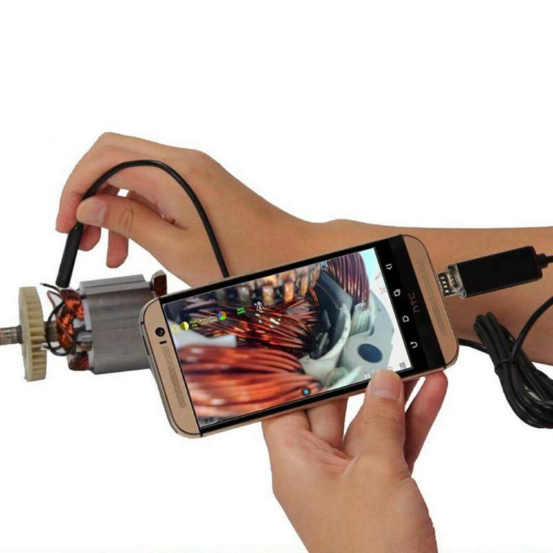
An endoscope is a medical device used to visualize the internal organs and structures of the body. It consists of a long, flexible tube with a light source and a camera at one end. The camera, known as an image sensor, plays a crucial role in capturing high-quality images or videos of the internal body parts.
The image sensor in an endoscope works by converting light into electrical signals. It is typically a charge-coupled device (CCD) or a complementary metal-oxide-semiconductor (CMOS) sensor. These sensors consist of an array of tiny light-sensitive pixels that detect the incoming light.
When the endoscope is inserted into the body, the light source illuminates the area of interest. The light reflects off the internal structures and enters the endoscope through a lens system. The lens focuses the light onto the image sensor, which then converts the light into electrical signals.
The electrical signals are then processed by the endoscope's electronics and transmitted to a monitor or a digital recording device. The monitor displays the real-time images or videos, allowing the healthcare professional to examine the internal body parts.
In recent years, there have been advancements in endoscope technology, particularly in the field of image sensors. The latest image sensors offer higher resolution, improved sensitivity to light, and better image quality. This allows for more accurate diagnosis and better visualization of the internal organs.
Additionally, some endoscopes now incorporate advanced features such as image stabilization and digital zoom, further enhancing the quality of the captured images. These advancements in image sensor technology have greatly improved the effectiveness and reliability of endoscopic procedures, leading to better patient outcomes.
In conclusion, the image sensor in an endoscope plays a crucial role in capturing high-quality images or videos of the internal body parts. Advancements in image sensor technology have significantly improved the visualization capabilities of endoscopes, leading to better diagnostic accuracy and patient care.













There are no comments for this blog.