How Does Endoscope Work ?
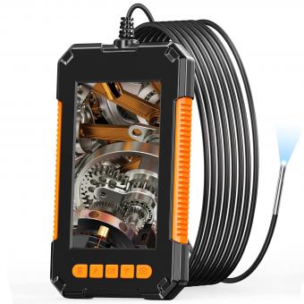
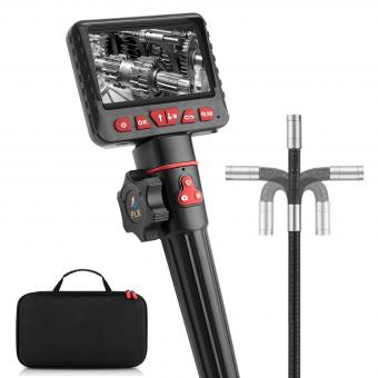
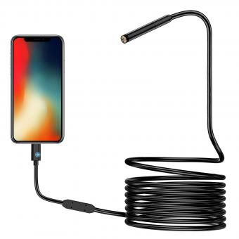
An endoscope is a medical device used to visualize and examine the interior of the body. It consists of a long, flexible tube with a light source and a camera at one end. The tube is inserted into the body through a natural opening or a small incision.
The endoscope works by transmitting light through fiber optic cables to illuminate the area being examined. The camera captures real-time images or videos, which are then displayed on a monitor for the healthcare professional to view. Some endoscopes also have channels that allow for the insertion of surgical instruments to perform procedures or collect samples.
The flexibility of the endoscope allows it to navigate through the body's internal structures, such as the digestive tract or respiratory system, providing a detailed visual examination. It is commonly used for diagnostic purposes, such as detecting abnormalities or diseases, and can also be used for therapeutic interventions, such as removing polyps or taking biopsies.
1、 Optical Fiber Transmission
An endoscope is a medical device that allows doctors to visualize and examine the internal organs and structures of the body. It consists of a long, flexible tube with a light source and a camera at one end. The other end is connected to a monitor, which displays the images captured by the camera.
The key component that enables the functioning of an endoscope is optical fiber transmission. Optical fibers are thin, flexible strands made of high-quality glass or plastic. They are designed to transmit light signals over long distances with minimal loss of intensity.
In the case of an endoscope, the light source at one end of the device emits a beam of light, which is then directed into the optical fiber. The light travels through the fiber, bouncing off the walls due to the principle of total internal reflection. This ensures that the light remains confined within the fiber and does not escape.
As the light reaches the other end of the fiber, it is directed towards the target area inside the body. The light illuminates the area, allowing the camera to capture clear and detailed images. These images are then transmitted back through the optical fiber to the monitor, where they are displayed in real-time for the doctor to observe and analyze.
The use of optical fiber transmission in endoscopes offers several advantages. Firstly, it allows for the transmission of light over long distances, enabling the endoscope to reach deep into the body. Secondly, the flexibility of the fibers allows for easy maneuverability within the body, minimizing discomfort for the patient. Lastly, the high-quality images produced by the camera provide valuable information for accurate diagnosis and treatment.
In recent years, there have been advancements in endoscope technology, such as the development of smaller and more flexible fibers. These advancements have further improved the functionality and effectiveness of endoscopes, allowing for better visualization and diagnosis of various medical conditions.
In conclusion, optical fiber transmission is a crucial component in the functioning of endoscopes. It enables the transmission of light from the source to the target area inside the body, allowing for clear visualization and examination. The continuous advancements in endoscope technology continue to enhance the capabilities of these devices, leading to improved medical outcomes.

2、 Illumination System
The illumination system is a crucial component of an endoscope that enables clear visualization of the internal body structures. It consists of various components that work together to provide adequate lighting during endoscopic procedures.
The primary element of the illumination system is the light source, which can be either a halogen or xenon lamp. These lamps emit intense light that is transmitted through a fiber optic cable to the distal end of the endoscope. The distal end contains a bundle of optical fibers that distribute the light evenly across the field of view.
The light emitted from the distal end illuminates the target area inside the body, allowing the endoscopist to visualize the tissues and organs. The reflected light is then captured by the objective lens and transmitted back through the endoscope to the eyepiece or camera system.
In recent years, there have been advancements in endoscope illumination systems. LED (Light Emitting Diode) technology has gained popularity due to its energy efficiency, longer lifespan, and compact size. LED lights provide consistent and bright illumination, enhancing the image quality during endoscopic procedures.
Furthermore, some endoscopes now incorporate advanced features such as adjustable light intensity and color temperature. These features allow the endoscopist to optimize the lighting conditions based on the specific requirements of the procedure and the target area.
In conclusion, the illumination system in an endoscope plays a vital role in providing adequate lighting for clear visualization during endoscopic procedures. With advancements in technology, the use of LED lights and adjustable lighting features has further improved the quality and versatility of endoscopic illumination systems.
3、 Lens System
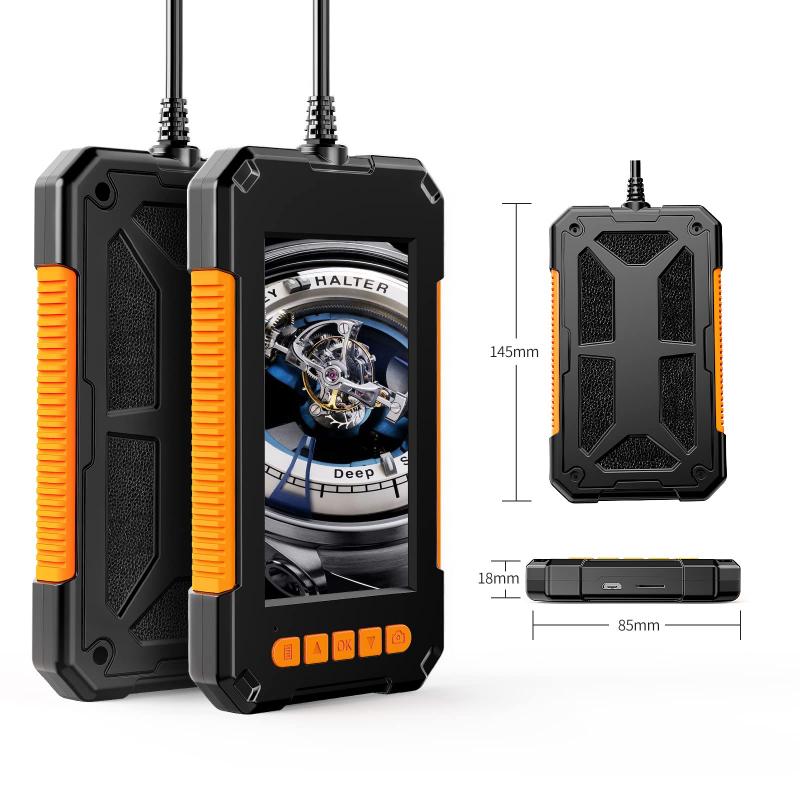
The endoscope is a medical device that allows doctors to visualize and examine the internal organs and structures of the body. It consists of a long, flexible tube with a light source and a camera at one end, and a lens system at the other end. The lens system is a crucial component of the endoscope as it helps to capture clear and detailed images of the internal body parts.
The lens system in an endoscope works by focusing light onto the camera sensor, which then transmits the images to a monitor for the doctor to view. The lens system is designed to provide a wide field of view and high-resolution images, allowing for accurate diagnosis and treatment.
The latest advancements in endoscope lens systems have focused on improving image quality and enhancing the overall performance of the device. One notable development is the use of high-definition cameras and advanced optics, which provide sharper and more detailed images. Additionally, some endoscopes now incorporate image stabilization technology to minimize motion blur and improve image clarity.
Another recent innovation is the integration of artificial intelligence (AI) algorithms into endoscope systems. AI can analyze the captured images in real-time, helping doctors to detect abnormalities or lesions that may be difficult to spot with the naked eye. This technology has the potential to improve diagnostic accuracy and assist in early detection of diseases.
In conclusion, the lens system in an endoscope plays a crucial role in capturing clear and detailed images of the internal body parts. The latest advancements in endoscope technology, such as high-definition cameras and AI integration, have further improved the image quality and diagnostic capabilities of these devices.
4、 Image Sensor
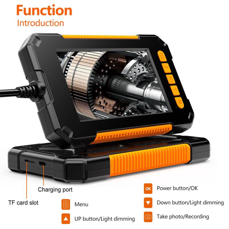
An endoscope is a medical device used to visualize the internal organs and structures of the body. It consists of a long, flexible tube with a light source and a camera at one end, and a viewing screen at the other end. The camera, also known as the image sensor, plays a crucial role in capturing high-quality images or videos of the internal body parts.
The image sensor in an endoscope works by converting light into electrical signals. It is typically a charge-coupled device (CCD) or a complementary metal-oxide-semiconductor (CMOS) sensor. These sensors consist of an array of tiny light-sensitive pixels that detect the incoming light.
When the endoscope is inserted into the body, the light source illuminates the area of interest. The light reflects off the internal structures and enters the camera through a lens system. The lens focuses the light onto the image sensor, which then converts the light into electrical signals.
The electrical signals are then processed by the endoscope's electronics and transmitted to the viewing screen. The screen displays the captured images or videos in real-time, allowing the medical professional to examine the internal structures and make a diagnosis.
In recent years, there have been advancements in endoscope technology, particularly in the field of image sensors. The latest image sensors offer higher resolution, improved sensitivity to light, and better image quality. This allows for more accurate and detailed visualization of the internal body parts, aiding in the diagnosis and treatment of various medical conditions.
Furthermore, some endoscopes now incorporate advanced features such as image stabilization and digital zoom, which further enhance the quality of the captured images. These advancements in image sensor technology continue to improve the effectiveness and reliability of endoscopic procedures, leading to better patient outcomes.
