How Does Light Microscope Work?
A light microscope works by using visible light to magnify and resolve small objects or structures that are otherwise too small to be seen by the naked eye. The light passes through a series of lenses, which refract and focus the light onto the specimen being observed. The specimen is placed on a glass slide and covered with a coverslip to protect it and keep it in place.
As the light passes through the specimen, it is absorbed, transmitted, or reflected depending on the properties of the specimen. The light that is transmitted through the specimen is then magnified again by the objective lens, which is located just below the specimen. The magnified image is then projected through the eyepiece, which further magnifies the image and allows the observer to see it.
The resolution of a light microscope is limited by the wavelength of visible light, which is around 400-700 nanometers. This means that it can only resolve structures that are larger than this size. However, by using different types of lenses and techniques such as staining, contrast enhancement, and fluorescence, it is possible to improve the resolution and visualize smaller structures.
1、 Magnification

How does light microscope work?
A light microscope works by using visible light to magnify small objects. The light passes through a series of lenses, which bend the light and focus it onto the object being viewed. The magnification of the microscope is determined by the combination of lenses used, with higher magnification achieved by using lenses with a shorter focal length.
The light microscope has been a fundamental tool in biology and medicine for centuries, allowing scientists to observe and study cells, tissues, and microorganisms. However, the resolution of light microscopes is limited by the wavelength of visible light, which is around 400-700 nanometers. This means that structures smaller than this cannot be resolved by a light microscope.
Recent advances in microscopy have led to the development of super-resolution techniques, such as stimulated emission depletion (STED) microscopy and structured illumination microscopy (SIM). These techniques use specialized optics and fluorescent dyes to overcome the diffraction limit of light and achieve resolutions of a few tens of nanometers.
In summary, a light microscope works by using visible light to magnify small objects, with the magnification determined by the combination of lenses used. While the resolution of light microscopes is limited by the wavelength of visible light, recent advances in microscopy have led to the development of super-resolution techniques that can overcome this limitation.
2、 Resolution

Resolution is one of the key concepts in understanding how a light microscope works. The resolution of a microscope refers to its ability to distinguish two closely spaced objects as separate entities. The resolution of a light microscope is limited by the wavelength of light used to illuminate the specimen. This is known as the diffraction limit, and it means that the smallest distance between two objects that can be resolved is approximately half the wavelength of the light used.
To improve the resolution of a light microscope, various techniques have been developed. One of the most important is the use of immersion oil, which has a refractive index similar to that of glass and reduces the amount of light that is scattered as it passes through the specimen. Another technique is the use of fluorescent dyes, which can be used to label specific structures within the specimen and make them more visible.
In recent years, there has been a growing interest in the development of super-resolution microscopy techniques, which allow for the visualization of structures that are smaller than the diffraction limit. These techniques include stimulated emission depletion (STED) microscopy, structured illumination microscopy (SIM), and single-molecule localization microscopy (SMLM). These techniques use various methods to overcome the diffraction limit, such as the use of special light sources or the detection of individual fluorescent molecules.
Overall, the light microscope works by using light to illuminate a specimen and magnifying the image using a series of lenses. The resolution of the microscope is limited by the wavelength of light used, but various techniques have been developed to improve the resolution and allow for the visualization of structures that are smaller than the diffraction limit.
3、 Illumination

Illumination is a crucial component of how a light microscope works. The microscope uses a light source, typically an LED or halogen bulb, to illuminate the sample being observed. The light passes through a condenser lens, which focuses the light onto the sample. The sample then scatters the light, and some of it enters the objective lens of the microscope. The objective lens magnifies the scattered light, and the image is then further magnified by the eyepiece lens, which the observer looks through.
In recent years, advancements in technology have allowed for even more precise and detailed imaging with light microscopes. One such advancement is the use of fluorescence microscopy, which involves labeling specific molecules or structures within the sample with fluorescent dyes. When illuminated with a specific wavelength of light, the labeled structures emit light of a different color, allowing for even greater contrast and specificity in imaging.
Another recent development is the use of super-resolution microscopy, which allows for imaging at a resolution beyond the diffraction limit of light. This is achieved through various techniques, such as stimulated emission depletion (STED) microscopy and structured illumination microscopy (SIM), which use complex light patterns to overcome the limitations of traditional microscopy.
Overall, the illumination component of a light microscope is essential for producing clear and detailed images of samples. Ongoing advancements in technology continue to push the boundaries of what is possible with light microscopy, allowing for even greater insights into the microscopic world.
4、 Lenses
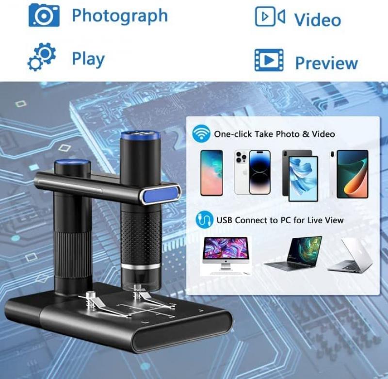
How does a light microscope work? The answer lies in the use of lenses. Light microscopes use a combination of lenses to magnify an object and allow us to see it in greater detail. The lenses work by bending the light that passes through them, which changes the direction of the light and makes the object appear larger.
The basic components of a light microscope include an objective lens, an eyepiece lens, and a light source. The objective lens is located near the specimen and is responsible for magnifying the image. The eyepiece lens is located near the viewer's eye and further magnifies the image. The light source illuminates the specimen, making it easier to see.
When light passes through the objective lens, it is refracted and focused onto the specimen. The light then passes through the specimen and is refracted again as it passes through the eyepiece lens. This creates a magnified image that the viewer can see.
Recent advancements in light microscopy have allowed for even greater magnification and resolution. Techniques such as confocal microscopy and super-resolution microscopy use specialized lenses and computer algorithms to create highly detailed images of cells and tissues. These techniques have revolutionized the field of biology and have allowed scientists to study the inner workings of cells in unprecedented detail.
In conclusion, light microscopes work by using lenses to bend and magnify light, allowing us to see objects in greater detail. Recent advancements in microscopy have allowed for even greater magnification and resolution, opening up new avenues of research in the field of biology.


