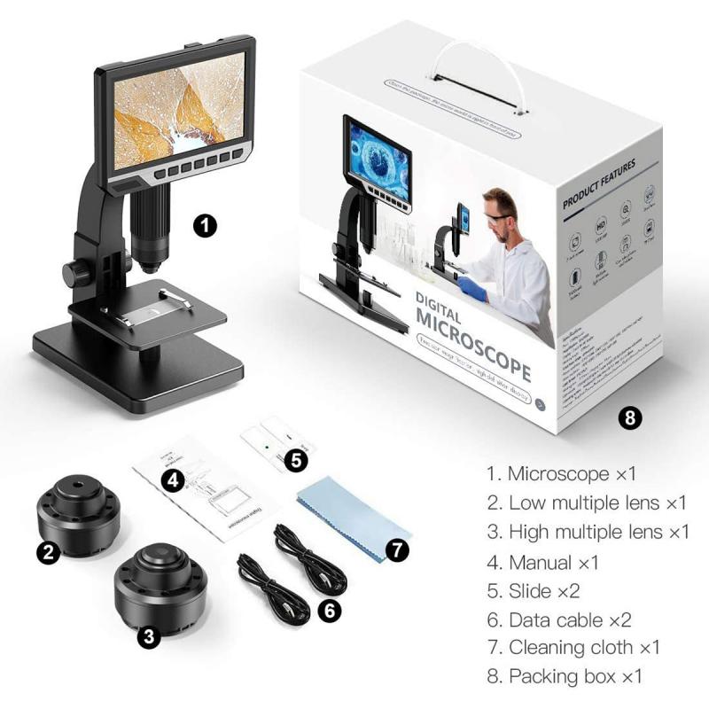
How Does Microscope Magnification Work ?
Microscope magnification works by using a combination of lenses to enlarge the image of a specimen. The objective lens, located near the specimen, produces a magnified image that is further magnified by the eyepiece lens. The total magnification is calculated by multiplying the magnification of the objective lens by the magnification of the eyepiece lens.
For example, if the objective lens has a magnification of 10x and the eyepiece lens has a magnification of 20x, the total magnification would be 200x (10 x 20 = 200).
The resolution of the microscope, or its ability to distinguish between two closely spaced objects, is also affected by magnification. As magnification increases, the resolution decreases, making it more difficult to distinguish fine details.
To overcome this limitation, microscopes often use special techniques such as oil immersion or electron microscopy to increase resolution and provide clearer images.
1、 Optical magnification
Optical magnification is the process by which a microscope enlarges an object to make it visible to the human eye. The magnification of a microscope is determined by the combination of lenses used in the instrument. The objective lens, located near the specimen, produces a magnified image that is further magnified by the eyepiece lens. The total magnification is calculated by multiplying the magnification of the objective lens by the magnification of the eyepiece lens.
The magnification of a microscope is limited by the wavelength of light used to illuminate the specimen. The maximum magnification that can be achieved with a light microscope is around 2000x. Beyond this limit, the image becomes blurry and distorted due to the diffraction of light. To overcome this limitation, electron microscopes use a beam of electrons instead of light to illuminate the specimen. This allows for much higher magnification, up to 10 million times, and greater resolution.
In recent years, advancements in technology have led to the development of super-resolution microscopy techniques, such as stimulated emission depletion (STED) microscopy and structured illumination microscopy (SIM). These techniques use specialized optics and fluorescent dyes to achieve resolutions beyond the diffraction limit of light. This has allowed scientists to study biological structures and processes at the molecular level, providing new insights into the workings of cells and tissues.

2、 Numerical aperture
Microscope magnification works by increasing the apparent size of an object being viewed through the microscope. This is achieved by using lenses to bend and focus light rays, which then pass through the object and into the microscope's eyepiece or camera. The magnification power of a microscope is determined by the combination of lenses used, and is typically expressed as a ratio of the size of the image seen through the microscope to the actual size of the object being viewed.
One important factor that affects microscope magnification is the numerical aperture (NA) of the lenses used. The NA is a measure of the ability of a lens to gather and focus light, and is determined by the refractive index of the lens material and the angle at which light enters the lens. Higher NA lenses are able to gather more light and produce sharper, more detailed images with greater magnification.
Recent advances in microscope technology have led to the development of super-resolution microscopy techniques, which allow for even higher levels of magnification and resolution than traditional microscopy methods. These techniques use specialized lenses and imaging methods to overcome the diffraction limit of light, which previously limited the resolution of optical microscopes. As a result, scientists are now able to study biological structures and processes at the molecular level, opening up new avenues for research and discovery.

3、 Resolution
Microscope magnification works by using lenses to bend and focus light, allowing the user to see objects that are too small to be seen with the naked eye. The magnification power of a microscope is determined by the combination of lenses used, with higher magnification achieved by using lenses with a shorter focal length.
However, magnification alone is not enough to produce a clear and detailed image. The ability of a microscope to distinguish between two closely spaced objects is known as its resolution. Resolution is determined by the wavelength of the light used and the numerical aperture of the lenses. The numerical aperture is a measure of the lens's ability to gather light and is determined by the lens's design and the refractive index of the medium between the lens and the specimen.
In recent years, there have been significant advancements in microscopy technology, including the development of super-resolution microscopy techniques. These techniques use specialized lenses and fluorescent dyes to overcome the diffraction limit of light, allowing for the visualization of structures at the nanoscale level.
Overall, microscope magnification and resolution are essential factors in producing clear and detailed images of microscopic structures. Advancements in technology continue to push the boundaries of what is possible in microscopy, allowing for new discoveries and insights into the world of the very small.

4、 Contrast
How does microscope magnification work? Microscope magnification works by using a combination of lenses to enlarge the image of a specimen. The objective lens, located near the specimen, produces a magnified image that is further magnified by the eyepiece lens. The total magnification is calculated by multiplying the magnification of the objective lens by the magnification of the eyepiece lens.
Contrast is another important factor in microscopy. Contrast refers to the difference in brightness between the specimen and its background. Without contrast, it would be difficult to distinguish the specimen from its surroundings. There are several ways to increase contrast in microscopy, including staining the specimen with dyes or using special lighting techniques.
In recent years, advances in microscopy technology have allowed for even greater magnification and resolution. Super-resolution microscopy techniques, such as stimulated emission depletion (STED) microscopy and structured illumination microscopy (SIM), have enabled researchers to visualize structures at the nanoscale level. These techniques use specialized equipment and software to overcome the limitations of traditional microscopy and provide unprecedented detail and clarity.
Overall, microscope magnification and contrast are essential components of microscopy that allow researchers to study the structure and function of biological specimens at various levels of detail. As technology continues to advance, we can expect even more exciting developments in the field of microscopy.


