How Does Optical Microscope Work ?
An optical microscope works by using visible light to magnify and observe small objects or specimens. It consists of several key components, including a light source, condenser lens, objective lens, eyepiece, and stage.
The light source, typically a bulb, emits light that passes through a condenser lens, which focuses the light onto the specimen. The objective lens, located near the specimen, further magnifies the image and collects the light that passes through it. The eyepiece, positioned at the top of the microscope, further magnifies the image and allows the viewer to observe it.
As light passes through the specimen, it interacts with the object, causing some of the light to be absorbed or scattered. The remaining light passes through the objective lens and forms an enlarged and inverted image. This image is then magnified again by the eyepiece, allowing the viewer to see a larger and more detailed version of the specimen.
By adjusting the focus and manipulating the lenses, the viewer can observe different parts of the specimen and study its structure, texture, and other characteristics.
1、 Principles of light propagation and refraction in optical microscopy.
The optical microscope is a widely used tool in scientific research and medical diagnostics. It works based on the principles of light propagation and refraction in optical microscopy.
When light passes through a sample, it undergoes several interactions, including absorption, scattering, and refraction. The optical microscope utilizes these interactions to produce an image of the sample.
In a basic optical microscope, a light source, typically a lamp, emits light that passes through a condenser lens. The condenser lens focuses the light onto the sample, which may be a thin section of tissue or a microscopic organism. The light interacts with the sample, and some of it is absorbed, while the rest is scattered or refracted.
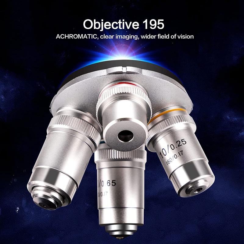
The scattered or refracted light then enters the objective lens, which collects and magnifies the light. The objective lens forms an enlarged real image of the sample, which is further magnified by the eyepiece lens. The observer views this magnified image through the eyepiece, allowing for detailed examination of the sample.
In recent years, advancements in optical microscopy have led to the development of various techniques that enhance the resolution and contrast of the images. For example, confocal microscopy uses a pinhole aperture to eliminate out-of-focus light, resulting in improved image quality and depth perception. Super-resolution microscopy techniques, such as stimulated emission depletion (STED) microscopy and structured illumination microscopy (SIM), have pushed the limits of resolution beyond the diffraction limit, enabling the visualization of subcellular structures and molecular interactions.
Overall, the optical microscope works by exploiting the principles of light propagation and refraction to produce magnified images of samples, and ongoing advancements continue to enhance its capabilities in various fields of research and diagnostics.
2、 Components and functions of an optical microscope.
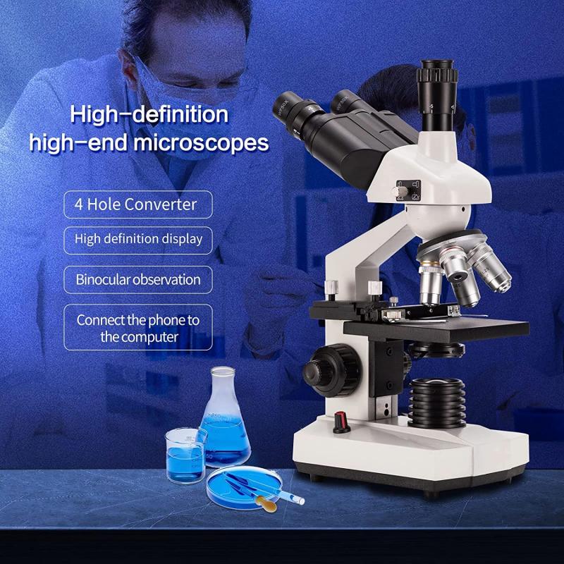
Components and functions of an optical microscope:
An optical microscope is a powerful tool used to magnify and observe small objects that are not visible to the naked eye. It consists of several key components that work together to produce a magnified image.
1. Objective lens: The objective lens is the primary lens of the microscope and is responsible for gathering light from the specimen. It has a high numerical aperture, which determines the resolution and clarity of the image.
2. Eyepiece lens: The eyepiece lens is located at the top of the microscope and is where the observer looks through to view the specimen. It further magnifies the image produced by the objective lens.
3. Stage: The stage is a flat platform where the specimen is placed for observation. It can be moved vertically and horizontally to position the specimen under the objective lens.
4. Illumination system: The illumination system provides light to illuminate the specimen. In most modern microscopes, this is achieved using an adjustable LED light source. The light passes through the specimen and is then collected by the objective lens.
5. Condenser lens: The condenser lens is located beneath the stage and focuses the light onto the specimen. It helps to improve the contrast and clarity of the image.
6. Focus adjustment: The focus adjustment mechanism allows the user to bring the specimen into sharp focus. This is typically achieved by moving the stage or adjusting the position of the objective lens.
The optical microscope works by using a combination of lenses and light to magnify and resolve the details of a specimen. When light passes through the specimen, it interacts with the structures and objects within it, causing some of the light to be absorbed or scattered. The remaining light is collected by the objective lens, which forms an enlarged and inverted image of the specimen. This image is then further magnified by the eyepiece lens, allowing the observer to see the details of the specimen.
In recent years, advancements in optical microscopy have led to the development of techniques such as confocal microscopy, which uses a pinhole to eliminate out-of-focus light and produce high-resolution images. Additionally, super-resolution microscopy techniques, such as stimulated emission depletion (STED) microscopy and structured illumination microscopy (SIM), have pushed the limits of optical resolution, allowing scientists to observe structures at the nanoscale.
Overall, the optical microscope remains a fundamental tool in scientific research, education, and various fields of study, providing valuable insights into the microscopic world.
3、 Illumination techniques in optical microscopy.
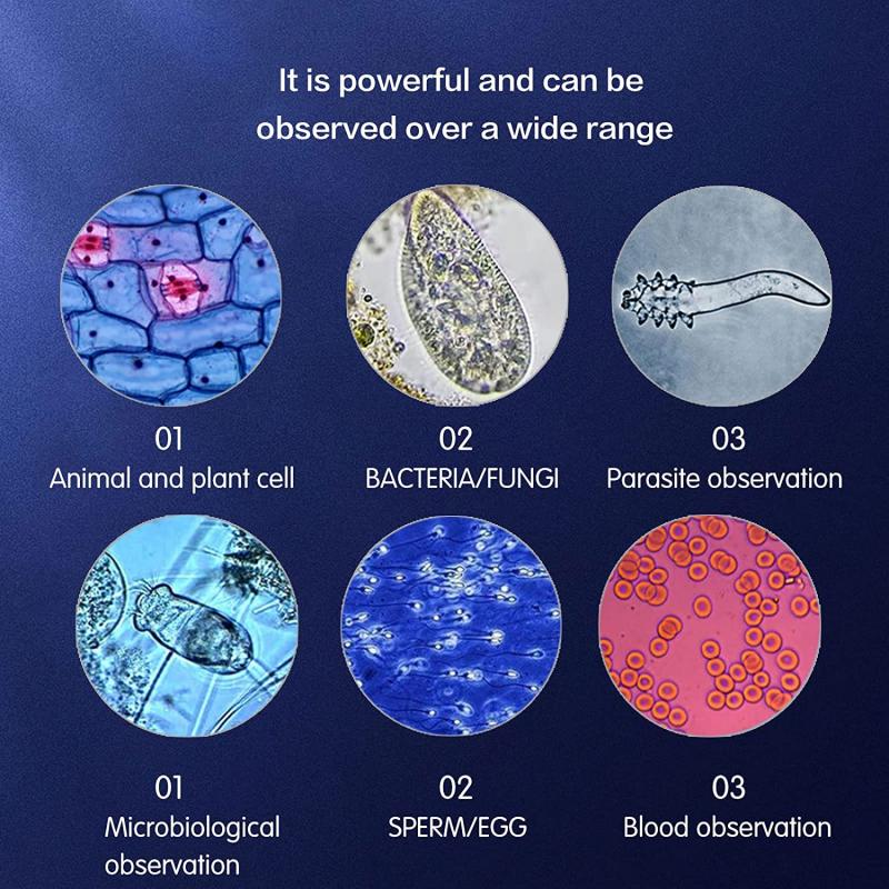
Illumination techniques in optical microscopy play a crucial role in enhancing the quality and resolution of images obtained through an optical microscope. These techniques involve the careful manipulation of light to illuminate the specimen being observed. By understanding how these techniques work, we can gain insights into the functioning of an optical microscope.
One of the primary illumination techniques used in optical microscopy is brightfield illumination. In this technique, a light source located beneath the specimen emits light, which passes through the condenser lens and is focused onto the specimen. The specimen absorbs some of the light, while the rest is transmitted through it. The objective lens then collects the transmitted light and forms an image that is magnified and observed by the eyepiece. Brightfield illumination is commonly used for observing stained or naturally pigmented specimens.
Another illumination technique is darkfield illumination, which involves placing a special condenser with an opaque disk in the light path. This disk blocks the direct light from reaching the objective lens, resulting in a dark background. However, light scattered by the specimen enters the objective lens, creating a bright image of the specimen against the dark background. Darkfield illumination is particularly useful for observing transparent or unstained specimens, as it enhances contrast and reveals fine details that may be otherwise difficult to see.
Fluorescence microscopy is another powerful illumination technique that utilizes fluorescent dyes or proteins to label specific structures within a specimen. The specimen is illuminated with a specific wavelength of light, which excites the fluorescent molecules, causing them to emit light of a different wavelength. This emitted light is then collected by the objective lens and observed. Fluorescence microscopy allows for the visualization of specific molecules or structures within cells and tissues, making it invaluable in various fields of research, including cell biology and immunology.
In recent years, advancements in illumination techniques have led to the development of super-resolution microscopy. This technique overcomes the diffraction limit of light, allowing for the visualization of structures at a resolution beyond what was previously thought possible. Techniques such as stimulated emission depletion (STED) microscopy and structured illumination microscopy (SIM) utilize complex illumination patterns and clever manipulation of light to achieve super-resolution imaging. These techniques have revolutionized the field of microscopy, enabling researchers to observe intricate details within cells and tissues that were previously inaccessible.
In conclusion, illumination techniques in optical microscopy are essential for enhancing image quality and resolution. Brightfield, darkfield, and fluorescence microscopy are commonly used techniques, each with its own advantages and applications. Furthermore, the recent advancements in super-resolution microscopy have pushed the boundaries of what can be observed using optical microscopy, opening up new avenues for scientific discovery.
4、 Image formation and magnification in optical microscopy.
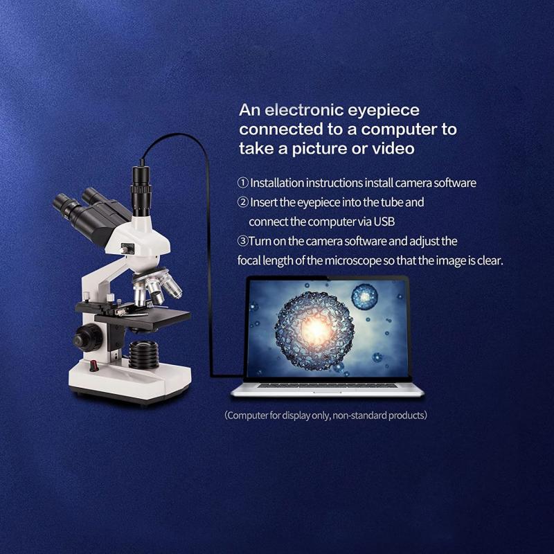
Optical microscopes work by using visible light to magnify and create an image of a sample. The basic principle behind their operation is the interaction of light with the sample and the subsequent formation of an image.
When light passes through the objective lens of an optical microscope, it is refracted and focused onto the sample. The sample interacts with the light, scattering and absorbing some of it. The scattered light then enters the objective lens again and is further refracted. This refracted light is collected by the eyepiece or camera, which forms a magnified image of the sample.
The magnification in optical microscopy is achieved through a combination of the objective lens and the eyepiece. The objective lens has a high numerical aperture, which determines its ability to gather light and resolve fine details. The eyepiece further magnifies the image formed by the objective lens, allowing the observer to see a larger and more detailed view of the sample.
In recent years, advancements in optical microscopy have led to the development of techniques such as confocal microscopy, multiphoton microscopy, and super-resolution microscopy. These techniques have pushed the limits of resolution and imaging capabilities, allowing scientists to observe structures and processes at the nanoscale.
Confocal microscopy uses a pinhole to eliminate out-of-focus light, resulting in improved image contrast and resolution. Multiphoton microscopy utilizes longer wavelength light and non-linear excitation to reduce photodamage and penetrate deeper into samples. Super-resolution microscopy techniques, such as stimulated emission depletion (STED) microscopy and structured illumination microscopy (SIM), surpass the diffraction limit of light, enabling the visualization of structures as small as a few nanometers.
Overall, optical microscopes continue to be a fundamental tool in scientific research, providing valuable insights into the microscopic world. The latest advancements in optical microscopy techniques have expanded our understanding of biological processes and materials science, opening up new avenues for exploration and discovery.
