How Fluorescent Microscope Works ?
A fluorescent microscope works by using a high-intensity light source to excite fluorescent molecules in a sample. The sample is first treated with a fluorescent dye or antibody that binds to specific molecules of interest. When the sample is illuminated with the light source, the fluorescent molecules absorb the light energy and emit light of a longer wavelength, which is then detected by the microscope's camera or eyepiece.
The emitted light is separated from the excitation light using filters, allowing only the fluorescent light to be visualized. This creates a highly contrasted image of the sample, with the fluorescent molecules appearing as bright spots against a dark background. Fluorescent microscopy is widely used in biological research to visualize specific molecules, such as proteins or DNA, within cells and tissues.
1、 Excitation light source

How fluorescent microscope works: Excitation light source
The excitation light source is a critical component of a fluorescent microscope. It is responsible for providing the energy required to excite the fluorescent molecules in the sample being observed. The excitation light source typically emits light in the ultraviolet or visible range, depending on the specific fluorescent molecule being used.
In traditional fluorescent microscopes, the excitation light source is typically a mercury or xenon arc lamp. However, in recent years, there has been a shift towards using LED-based excitation light sources. LED-based excitation light sources offer several advantages over traditional arc lamps, including longer lifetimes, lower power consumption, and improved stability.
When the excitation light source is turned on, it emits light that passes through the microscope's objective lens and illuminates the sample being observed. If the sample contains fluorescent molecules, the excitation light will cause these molecules to become excited and emit light of a longer wavelength. This emitted light is then collected by the microscope's objective lens and passed through a series of filters that separate the emitted light from the excitation light.
The emitted light is then focused onto a detector, such as a camera or photomultiplier tube, which converts the light into an electrical signal that can be processed and displayed as an image.
In summary, the excitation light source is a critical component of a fluorescent microscope, providing the energy required to excite fluorescent molecules in the sample being observed. LED-based excitation light sources offer several advantages over traditional arc lamps, including longer lifetimes, lower power consumption, and improved stability.
2、 Fluorescent dyes

Fluorescent microscopy is a powerful tool used in biological research to visualize and study cellular structures and processes. The technique relies on the use of fluorescent dyes, which are molecules that absorb light at a specific wavelength and emit light at a longer wavelength. This property allows them to be used to selectively label specific structures or molecules within cells.
When a sample is illuminated with light of the appropriate wavelength, the fluorescent dye molecules within the sample absorb the light and become excited. As the excited molecules return to their ground state, they emit light at a longer wavelength, which can be detected and visualized using a fluorescent microscope.
Fluorescent dyes can be used to label a wide range of cellular structures and molecules, including proteins, DNA, and organelles such as mitochondria and lysosomes. By selectively labeling specific structures or molecules, researchers can visualize and study their behavior and interactions within cells.
Recent advances in fluorescent microscopy have led to the development of super-resolution techniques, which allow for even higher resolution imaging of cellular structures and processes. These techniques use specialized fluorescent dyes and imaging methods to overcome the diffraction limit of traditional microscopy, allowing for imaging at the nanoscale level.
Overall, fluorescent microscopy and the use of fluorescent dyes have revolutionized the field of biological research, allowing for detailed visualization and study of cellular structures and processes.
3、 Filters
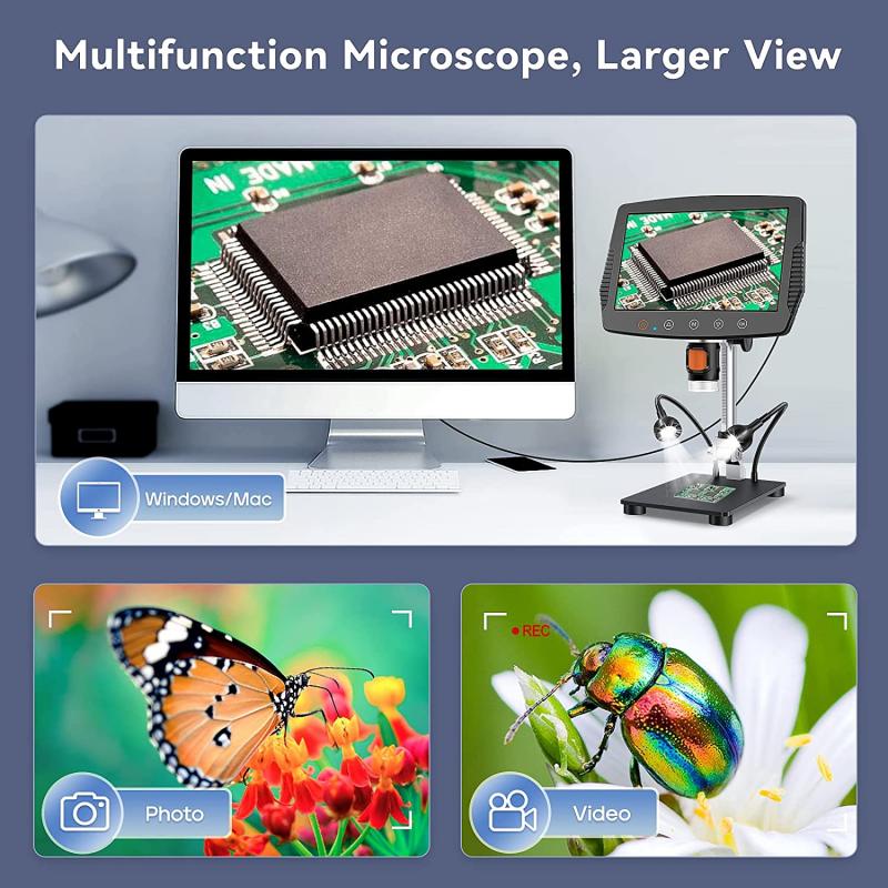
Filters are an essential component of a fluorescent microscope, which works by exciting fluorescent molecules in a sample with a specific wavelength of light and then detecting the emitted light at a different wavelength. Filters are used to select the excitation and emission wavelengths and to block unwanted light.
In a typical fluorescent microscope, a light source such as a mercury or xenon arc lamp provides the excitation light, which is directed through a filter that selects the desired wavelength. The excitation light then passes through the objective lens and illuminates the sample, causing the fluorescent molecules to emit light at a longer wavelength. This emitted light is collected by the same objective lens and passes through another filter that blocks the excitation light and selects the desired emission wavelength. The emitted light is then detected by a camera or photomultiplier tube.
Recent advances in filter technology have led to the development of more precise and efficient filters for fluorescent microscopy. For example, multiband filters can select multiple excitation and emission wavelengths simultaneously, allowing for more complex imaging experiments. Additionally, new types of filters such as notch filters and polarizing filters can further reduce unwanted light and improve image contrast.
Overall, filters are a critical component of fluorescent microscopy, allowing researchers to selectively excite and detect fluorescent molecules in a sample with high sensitivity and specificity.
4、 Objective lens

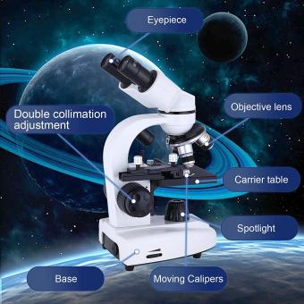
Objective lens is a crucial component of a fluorescent microscope that plays a vital role in producing high-resolution images of fluorescently labeled specimens. The objective lens is responsible for collecting and focusing the light emitted by the fluorescent sample onto the detector, which then produces an image.
The objective lens is designed to have a high numerical aperture (NA), which determines the amount of light that can be collected and the resolution of the image. The higher the NA, the better the resolution of the image. The objective lens is also designed to have a long working distance, which allows for the imaging of thick specimens.
In a fluorescent microscope, the objective lens is used in conjunction with a light source that excites the fluorescent molecules in the sample. The excited molecules emit light at a longer wavelength, which is then collected and focused by the objective lens.
Recent advancements in objective lens technology have led to the development of super-resolution microscopy techniques, such as stimulated emission depletion (STED) microscopy and structured illumination microscopy (SIM). These techniques use specialized objective lenses that can achieve resolutions beyond the diffraction limit of light, allowing for the imaging of structures at the nanoscale level.
In conclusion, the objective lens is a critical component of a fluorescent microscope that enables high-resolution imaging of fluorescently labeled specimens. Recent advancements in objective lens technology have led to the development of super-resolution microscopy techniques, which have revolutionized the field of microscopy.


