How Is Endoscopic Ultrasound Performed ?
Endoscopic ultrasound (EUS) is performed using a specialized endoscope equipped with an ultrasound probe at its tip. The procedure is typically done under sedation. The endoscope is inserted through the mouth or anus, depending on the area being examined.
During EUS, the ultrasound probe emits sound waves that create detailed images of the organs and tissues surrounding the gastrointestinal tract. These images help in diagnosing and staging various conditions, such as cancers, pancreatitis, and gastrointestinal disorders.
In addition to imaging, EUS allows for the collection of tissue samples (biopsy) or fluid aspiration for further analysis. This is done by inserting a needle through the endoscope and into the targeted area. EUS-guided interventions, such as drainage of fluid collections or injection of therapeutic agents, can also be performed.
Overall, EUS is a minimally invasive procedure that provides valuable diagnostic and therapeutic information, aiding in the management of various gastrointestinal conditions.
1、 Equipment and Setup for Endoscopic Ultrasound Procedure
Endoscopic ultrasound (EUS) is a minimally invasive procedure that combines endoscopy and ultrasound imaging to visualize and evaluate the gastrointestinal tract and surrounding structures. It is commonly used for diagnosing and staging various gastrointestinal diseases, including cancers.
How is endoscopic ultrasound performed?
1. Preparation: Before the procedure, the patient is typically given a sedative to help them relax. They may also receive a local anesthetic to numb the throat. The patient is positioned on their left side, and a mouthguard is placed to protect the teeth and endoscope.
2. Insertion of the endoscope: A thin, flexible tube called an endoscope is inserted through the mouth and guided down the esophagus into the stomach and duodenum. The endoscope has a light and a camera at its tip, allowing the doctor to see the internal structures.
3. Ultrasound imaging: Once the endoscope is in position, a small ultrasound probe is passed through a channel in the endoscope. The probe emits high-frequency sound waves that bounce off the organs and tissues, creating detailed images in real-time. The ultrasound images are displayed on a monitor, allowing the doctor to visualize the area of interest.
4. Fine-needle aspiration (FNA): In some cases, a fine needle may be passed through the endoscope to obtain tissue samples for further analysis. This is known as fine-needle aspiration (FNA) and can help in the diagnosis of tumors or other abnormalities.
5. Completion and recovery: Once the procedure is complete, the endoscope is slowly withdrawn. The patient is monitored until the sedative wears off, and they can resume normal activities within a few hours.
Equipment and Setup for Endoscopic Ultrasound Procedure:
The equipment used for EUS includes an endoscope with an ultrasound probe, a monitor to display the ultrasound images, and a fine needle for FNA. The endoscope is typically a flexible instrument with a diameter of 9-12 mm, allowing it to be easily maneuvered through the gastrointestinal tract. The ultrasound probe is usually located at the tip of the endoscope and can provide high-resolution images.
In recent years, there have been advancements in EUS technology. One notable development is the introduction of radial scanning ultrasound probes, which provide a 360-degree view of the surrounding tissues. This allows for better visualization and characterization of lesions. Additionally, there have been improvements in image resolution and the ability to perform elastography, which assesses tissue stiffness and can aid in the diagnosis of certain conditions.
The setup for an EUS procedure involves a dedicated endoscopy suite equipped with the necessary instruments and monitoring devices. The room is typically well-lit and has a comfortable environment for both the patient and the medical staff. Proper sterilization and infection control measures are followed to ensure patient safety.
In conclusion, endoscopic ultrasound is performed by inserting an endoscope into the gastrointestinal tract and using an ultrasound probe to obtain detailed images. The procedure requires specialized equipment and a dedicated setup. Ongoing advancements in EUS technology continue to improve the diagnostic capabilities and patient outcomes.
2、 Sedation and Patient Preparation for Endoscopic Ultrasound Examination
Endoscopic ultrasound (EUS) is a minimally invasive procedure that combines endoscopy and ultrasound to visualize and evaluate the gastrointestinal tract and adjacent structures. It is commonly used for diagnosing and staging various gastrointestinal diseases, including pancreatic cancer, gastrointestinal tumors, and gallbladder diseases.
To perform an endoscopic ultrasound, the patient is typically placed under sedation to ensure comfort and minimize any discomfort during the procedure. Sedation is usually administered intravenously by an anesthesiologist or nurse anesthetist. The level of sedation can vary depending on the patient's needs and the complexity of the procedure.
Patient preparation for an endoscopic ultrasound examination involves fasting for a certain period before the procedure. This is typically done to ensure that the stomach and duodenum are empty, allowing for better visualization of the target area. The fasting period may range from 6 to 12 hours, depending on the specific instructions provided by the healthcare provider.
In recent years, there have been advancements in sedation techniques for endoscopic ultrasound. The use of propofol, a short-acting sedative, has gained popularity due to its rapid onset and quick recovery time. Propofol allows for deeper sedation, which can be advantageous for complex procedures or patients who may be anxious or uncomfortable.
During the procedure, a thin, flexible endoscope with an ultrasound probe attached to its tip is inserted through the mouth or anus, depending on the area being examined. The ultrasound probe emits high-frequency sound waves that create detailed images of the gastrointestinal tract and surrounding structures. These images are then displayed on a monitor, allowing the healthcare provider to assess the condition of the organs and take biopsies if necessary.
In conclusion, endoscopic ultrasound is performed under sedation to ensure patient comfort. Patient preparation involves fasting, and advancements in sedation techniques, such as the use of propofol, have improved the overall experience for patients undergoing this procedure. The combination of endoscopy and ultrasound provides valuable diagnostic information and aids in the management of various gastrointestinal diseases.
3、 Insertion and Maneuvering of the Endoscope during Ultrasound Imaging
Endoscopic ultrasound (EUS) is a minimally invasive procedure that combines endoscopy and ultrasound imaging to visualize and evaluate the gastrointestinal tract and surrounding structures. It is commonly used for diagnosing and staging various gastrointestinal diseases, including cancers.
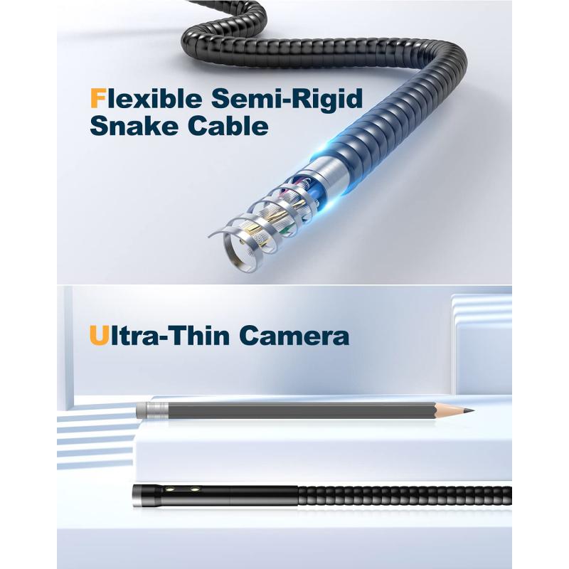
The procedure begins with the patient being sedated to ensure comfort. A thin, flexible endoscope with an ultrasound probe attached to its tip is inserted through the mouth or anus, depending on the area being examined. The endoscope allows the physician to see the inside of the gastrointestinal tract, while the ultrasound probe emits sound waves that create detailed images of the surrounding organs and tissues.
During the procedure, the endoscope is maneuvered to different positions to obtain comprehensive images. The physician can control the direction and depth of the ultrasound probe to visualize specific areas of interest. This allows for precise evaluation of the gastrointestinal wall, lymph nodes, blood vessels, and adjacent organs.
Advancements in technology have improved the maneuverability and image quality of EUS. The latest endoscopes have features like radial scanning and linear scanning. Radial scanning provides a 360-degree view of the surrounding tissues, while linear scanning allows for better visualization of structures in a straight line.
In recent years, there has been a growing interest in using EUS for therapeutic purposes, such as fine-needle aspiration (FNA) or biopsy. FNA involves using a needle attached to the endoscope to collect tissue samples for further analysis. This technique has proven to be highly accurate in diagnosing and staging gastrointestinal cancers.
Overall, EUS is a valuable tool in the diagnosis and management of gastrointestinal diseases. Its ability to provide detailed imaging and perform targeted biopsies makes it an essential procedure in the field of gastroenterology.
4、 Image Acquisition and Interpretation in Endoscopic Ultrasound
Endoscopic ultrasound (EUS) is a minimally invasive procedure that combines endoscopy and ultrasound imaging to visualize and evaluate the gastrointestinal tract and surrounding structures. It is commonly used for diagnosing and staging various gastrointestinal diseases, including pancreatic cancer, gastrointestinal tumors, and gallbladder diseases.
During an EUS procedure, a thin, flexible endoscope with an ultrasound probe attached to its tip is inserted through the mouth or anus and advanced into the esophagus, stomach, or rectum, depending on the area of interest. The ultrasound probe emits high-frequency sound waves that create detailed images of the organs and tissues in real-time.
To perform EUS, the patient is usually sedated to ensure comfort during the procedure. The endoscope is then carefully maneuvered to the desired location, and the ultrasound probe is used to obtain images from different angles. The images are displayed on a monitor, allowing the physician to visualize the structures and identify any abnormalities.
In recent years, there have been advancements in EUS technology, such as the development of high-frequency ultrasound probes and the addition of Doppler imaging. High-frequency probes provide better resolution and allow for more detailed imaging of superficial structures. Doppler imaging helps assess blood flow within the organs, aiding in the diagnosis of vascular abnormalities and guiding interventions.
After the images are acquired, the physician interprets them to make a diagnosis or determine the stage of a disease. EUS-guided fine-needle aspiration (EUS-FNA) or biopsy may also be performed during the procedure to obtain tissue samples for further analysis.
Overall, EUS is a valuable tool in the field of gastroenterology, providing detailed imaging and aiding in the diagnosis and management of various gastrointestinal diseases. The continuous advancements in technology further enhance its capabilities and improve patient outcomes.
