How To Check Sperm In Microscope ?
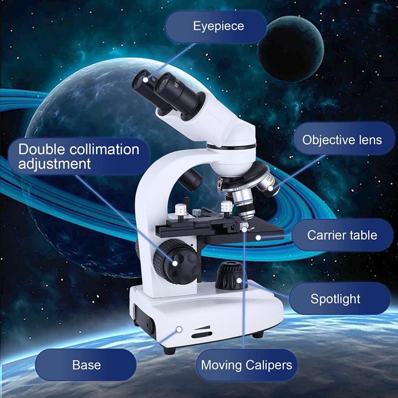
To check sperm under a microscope, you would first need to collect a semen sample. This can be done through masturbation or using a special condom during sexual intercourse. Once you have the sample, you would place a small drop onto a glass slide and cover it with a coverslip. The slide can then be placed under a microscope with a high magnification objective lens. By adjusting the focus and using proper lighting, you should be able to observe the sperm cells. It is important to note that proper training and knowledge of microscopy techniques are necessary to accurately identify and assess sperm morphology and motility.
1、 Sperm morphology: Assessing the size and shape of sperm cells.
To check sperm in a microscope and assess sperm morphology, follow these steps:
1. Collect a semen sample: The sample can be obtained through masturbation or by using a special condom during sexual intercourse. It is important to collect the sample in a sterile container to avoid contamination.
2. Prepare a slide: Take a clean glass slide and place a small drop of the semen sample onto it. Cover the drop with a coverslip to prevent drying.
3. Observe under a microscope: Place the slide on the stage of a microscope and start with a low magnification objective lens (e.g., 10x). Locate the area where the sperm cells are concentrated.
4. Increase magnification: Switch to a higher magnification objective lens (e.g., 40x or 100x) to observe the sperm cells in more detail. Adjust the focus until the sperm cells come into clear view.
5. Assess sperm morphology: Look for abnormalities in size and shape of the sperm cells. Normal sperm cells have an oval-shaped head, a long tail, and a smooth appearance. Abnormalities may include misshapen heads, double heads, or coiled tails.
It is worth noting that assessing sperm morphology alone may not provide a complete picture of male fertility. Other factors such as sperm count, motility, and overall sperm health should also be considered. Additionally, it is recommended to consult a healthcare professional or a fertility specialist for a comprehensive evaluation of male fertility. They may use advanced techniques like computer-assisted sperm analysis (CASA) to provide a more accurate assessment of sperm morphology and other parameters.
2、 Sperm motility: Evaluating the movement and swimming ability of sperm.
To check sperm in a microscope, you will need a few essential tools and follow specific steps. Here is a general guide on how to check sperm using a microscope:
1. Prepare the sample: Collect a semen sample in a sterile container through masturbation. It is important to abstain from ejaculation for 2-5 days before collecting the sample for accurate results.
2. Prepare the slide: Take a clean glass microscope slide and place a small drop of the semen sample onto it. Cover the drop with a coverslip to prevent drying.
3. Observe under the microscope: Place the slide on the microscope stage and start with a low magnification objective lens (e.g., 10x). Gradually increase the magnification to observe the sperm in detail.
4. Evaluate sperm motility: Sperm motility refers to the movement and swimming ability of sperm. Assess the percentage of motile sperm by counting the number of moving sperm in several fields of view. The World Health Organization (WHO) provides guidelines for classifying sperm motility into different categories, such as progressive motility (forward movement) and non-progressive motility (limited movement).
5. Assess sperm morphology: While observing the sperm, also evaluate their shape and structure. Abnormalities in sperm morphology can affect fertility. The WHO provides criteria for classifying normal and abnormal sperm morphology.
It is important to note that evaluating sperm under a microscope provides only a basic assessment of sperm quality. For a comprehensive analysis, it is recommended to consult a fertility specialist who can perform additional tests, such as semen analysis, to evaluate various parameters like sperm count, volume, pH, and more.
It is worth mentioning that advancements in technology have led to the development of computer-assisted sperm analysis (CASA) systems. These systems use specialized software to analyze sperm parameters automatically, providing more accurate and objective results. However, CASA systems are typically found in specialized laboratories and may not be readily available for individual use.
3、 Sperm count: Determining the concentration of sperm in a given sample.
To check sperm in a microscope and determine the sperm count, follow these steps:
1. Collect a semen sample: The sample should be collected through masturbation into a clean, sterile container. It is important to avoid any lubricants or condoms that may interfere with the accuracy of the test.
2. Allow the sample to liquefy: Semen typically becomes gel-like after ejaculation, but it will liquefy within 20-30 minutes at room temperature. This step is crucial for accurate analysis.
3. Prepare a microscope slide: Place a drop of the liquefied semen onto a clean glass microscope slide. Cover it with a coverslip to prevent drying.
4. Examine the sample under a microscope: Use a microscope with a magnification of at least 400x. Observe the slide under low power to locate an area where sperm are evenly distributed.
5. Count the sperm: Switch to high power and count the number of sperm in several fields of view. To determine the concentration, multiply the average number of sperm counted by the dilution factor (if applicable) and the volume of the sample.
It is worth noting that recent advancements in technology have introduced automated sperm analysis systems. These systems use computer algorithms to analyze sperm concentration, motility, and morphology. They provide more accurate and consistent results compared to manual methods, reducing human error and subjectivity.
However, it is important to consult a healthcare professional for a comprehensive evaluation of sperm health. They can provide a more detailed analysis, including factors like sperm motility and morphology, which are crucial for assessing fertility potential.
4、 Sperm vitality: Assessing the percentage of live sperm in a sample.
To check sperm in a microscope and assess sperm vitality, follow these steps:
1. Collect a semen sample: The sample should be collected through masturbation into a clean, sterile container. It is important to avoid any lubricants or condoms that may interfere with the accuracy of the test.
2. Prepare a slide: Take a small drop of the semen sample and place it on a clean glass slide. Add a coverslip gently to avoid air bubbles.
3. Observe under a microscope: Place the slide on the microscope stage and start with a low magnification objective lens. Locate the sperm cells on the slide.
4. Assess sperm vitality: To determine the percentage of live sperm, observe the sperm cells under high magnification. Live sperm will exhibit rapid, progressive movement, while dead sperm will be immobile or show slow, irregular movement.
5. Count and calculate: Count the number of live and dead sperm cells in several fields of view. Calculate the percentage of live sperm by dividing the number of live sperm by the total number of sperm observed and multiplying by 100.
It is worth noting that the assessment of sperm vitality through microscopy is a subjective method and can vary depending on the observer's experience. Additionally, recent advancements in technology have introduced automated sperm analysis systems that provide more accurate and objective results. These systems use computer algorithms to analyze sperm movement and viability, reducing human error and providing more reliable data.
However, manual microscopy remains a widely used method due to its accessibility and cost-effectiveness. It is important to consult with a healthcare professional or fertility specialist for a comprehensive evaluation of sperm health and fertility potential.
