How To Draw A Microscope And Label ?
To draw a microscope and label it, follow these steps:
1. Draw a rectangle for the base of the microscope.
2. Draw a vertical rectangle on top of the base for the body of the microscope.
3. Draw a smaller rectangle on top of the body for the eyepiece.
4. Draw a diagonal rectangle connecting the body and the eyepiece for the arm of the microscope.
5. Draw a circular shape on the arm for the objective lens.
6. Draw a smaller circular shape on the objective lens for the stage.
7. Draw two small rectangles on the stage for the clips that hold the slide in place.
8. Label the eyepiece as "ocular lens" and the objective lens as "objective lens".
9. Label the clips as "slide clips" and the stage as "stage".
10. Add any additional details or shading as desired.
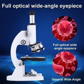
Your final drawing should resemble a typical compound microscope with all the necessary parts labeled for identification.
1、 Microscope Anatomy
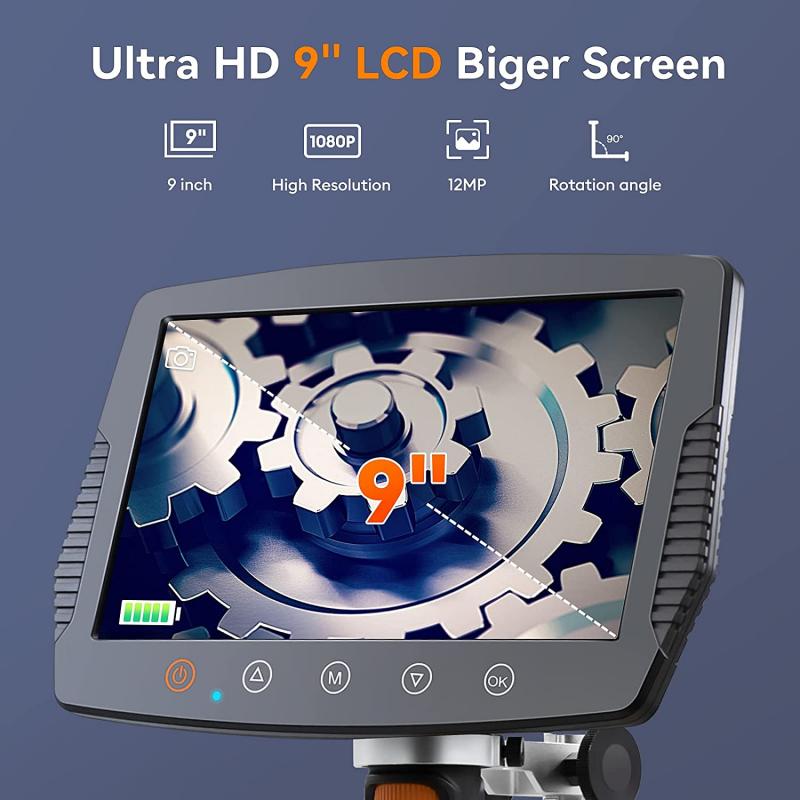
To draw a microscope and label its anatomy, follow these steps:
1. Draw the base of the microscope, which is the flat surface that supports the entire instrument.
2. Draw the arm of the microscope, which is the vertical part that connects the base to the head.
3. Draw the head of the microscope, which contains the eyepiece and the objective lenses.
4. Draw the eyepiece, which is the lens that you look through to view the specimen.
5. Draw the objective lenses, which are the lenses that magnify the specimen.
6. Label the stage, which is the flat platform where the specimen is placed.
7. Label the diaphragm, which is the part that controls the amount of light that passes through the specimen.
8. Label the focus knobs, which are used to adjust the focus of the microscope.
9. Label the coarse focus knob, which is used to make large adjustments to the focus.
10. Label the fine focus knob, which is used to make small adjustments to the focus.
11. Label the light source, which is usually located at the base of the microscope.
12. Label the power switch, which turns the microscope on and off.
It is important to note that there are different types of microscopes, such as compound microscopes and electron microscopes, which have different anatomy and functions. Additionally, advancements in technology have led to the development of digital microscopes, which allow for the capture and analysis of digital images of specimens.
2、 Labeling the Parts
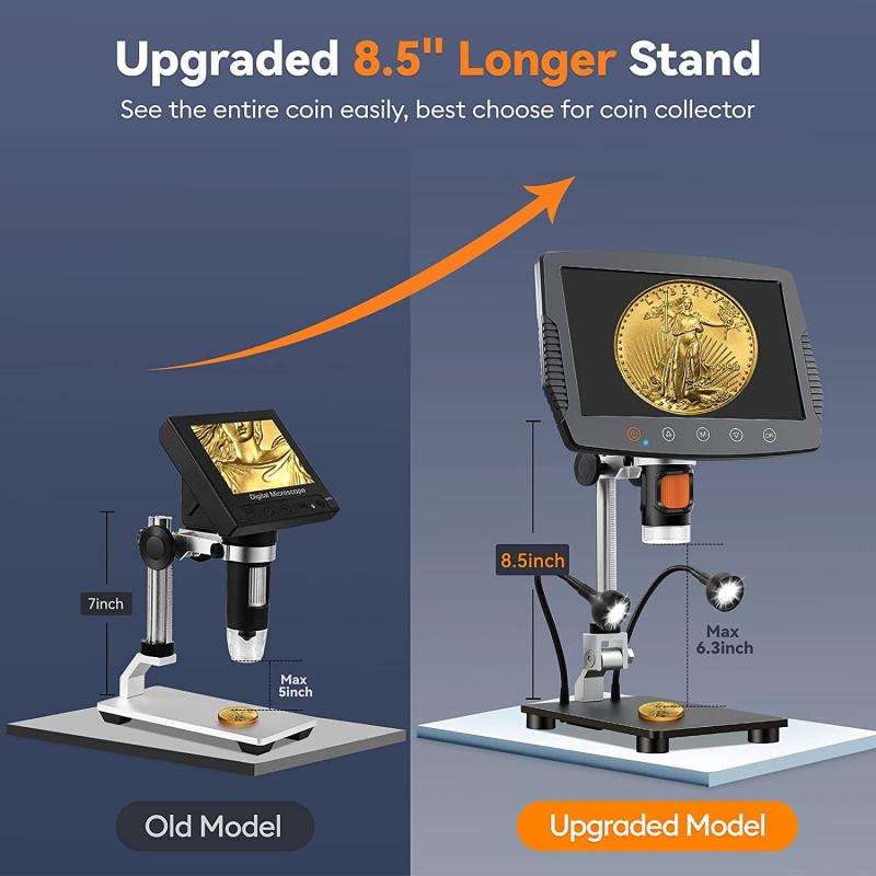
How to Draw a Microscope and Labeling the Parts
Drawing a microscope and labeling its parts is a great way to learn about the different components of this important scientific tool. Here are the steps to follow:
Step 1: Draw the basic shape of the microscope. Start by drawing a rectangle for the base and a smaller rectangle for the stage. Then draw a long, thin rectangle for the arm and a circular shape for the eyepiece.
Step 2: Add the lenses. Draw a larger circular shape for the objective lens and a smaller circular shape for the eyepiece lens. Connect the two lenses with a tube.
Step 3: Label the parts. Use arrows and labels to identify the different parts of the microscope. The base should be labeled "base," the stage should be labeled "stage," the arm should be labeled "arm," the eyepiece should be labeled "eyepiece," the objective lens should be labeled "objective lens," and the tube connecting the lenses should be labeled "tube."
Step 4: Add additional labels. Depending on the type of microscope you are drawing, you may need to add additional labels for other parts such as the diaphragm, the focus knobs, and the light source.
It's important to note that microscopes have evolved over time, and different types of microscopes may have different parts and features. For example, electron microscopes use a beam of electrons instead of light to magnify objects, and may have different components than a traditional light microscope. Therefore, it's important to research the specific type of microscope you are drawing to ensure accuracy in labeling its parts.
3、 Objective Lenses
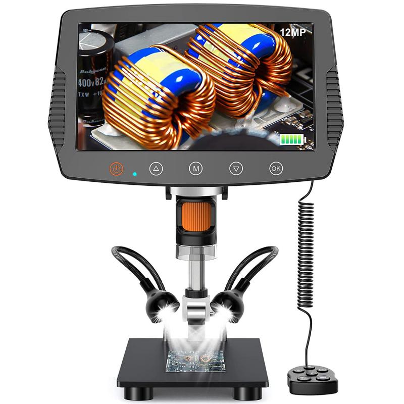
To draw a microscope and label the objective lenses, follow these steps:
1. Draw the base of the microscope as a rectangle with rounded corners.
2. Draw the arm of the microscope extending from the back of the base and curving upwards.
3. Draw the eyepiece at the top of the arm as a circle with a smaller circle inside.
4. Draw the objective lenses at the bottom of the arm as three circles of different sizes.
5. Label the objective lenses as "Low Power", "High Power", and "Oil Immersion" from left to right.
6. Add any additional details, such as the focus knobs and stage.
When labeling the objective lenses, it is important to note their magnification power and numerical aperture (NA). The Low Power lens typically has a magnification of 10x and an NA of 0.25, while the High Power lens has a magnification of 40x-100x and an NA of 0.65-0.85. The Oil Immersion lens has a magnification of 100x-120x and an NA of 1.25-1.4. It is important to use immersion oil with this lens to improve resolution.
It is also worth noting that modern microscopes may have additional objective lenses, such as a Super High Power lens with a magnification of 100x-150x and an NA of 1.45-1.6. These lenses are used for advanced microscopy techniques such as fluorescence and confocal microscopy.
Overall, drawing and labeling a microscope with objective lenses requires attention to detail and an understanding of their magnification and numerical aperture.
4、 Eyepiece and Magnification
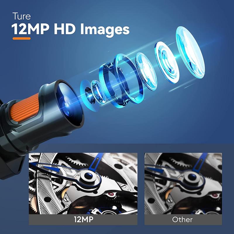
To draw a microscope and label the eyepiece and magnification, follow these steps:
1. Draw a rectangle for the base of the microscope.
2. Draw a vertical rectangle on top of the base for the body of the microscope.
3. Draw a smaller rectangle on top of the body for the eyepiece.
4. Draw a circle on the body for the objective lens.
5. Draw a diagonal line connecting the eyepiece to the objective lens.
6. Label the eyepiece as "ocular lens" and the objective lens as "objective lens".
7. Label the magnification of the eyepiece and objective lens, such as "10x" and "40x", respectively.
It's important to note that there are different types of microscopes with varying magnifications and features. For example, some microscopes have multiple objective lenses with different magnifications that can be switched out, while others have digital imaging capabilities. Additionally, advancements in technology have led to the development of more powerful microscopes, such as electron microscopes, which can magnify objects up to millions of times. As such, it's important to consider the specific type of microscope being drawn and labeled.


