How To Estimate Size Of Specimen Under Microscope ?
To estimate the size of a specimen under a microscope, you can use a technique called "micrometry." Micrometry involves measuring the size of the specimen using a calibrated eyepiece reticle or a stage micrometer.
First, place the specimen on the microscope stage and focus on it using the appropriate magnification. Then, switch to a lower magnification objective lens to have a wider field of view.
Next, place the calibrated eyepiece reticle or stage micrometer in the microscope's eyepiece. The reticle or micrometer should have a known scale, such as millimeters or micrometers, which can be used for measurement.
Align the scale on the reticle or micrometer with the scale of the specimen you want to measure. Count the number of divisions on the reticle or micrometer that span the length or width of the specimen.
Finally, multiply the number of divisions by the known scale value of the reticle or micrometer to calculate the size of the specimen. Make sure to take into account the magnification of the objective lens used during measurement.
1、 Measurement techniques for estimating specimen size under a microscope
To estimate the size of a specimen under a microscope, there are several measurement techniques that can be employed. These techniques involve the use of a calibrated eyepiece reticle or a stage micrometer, which allows for accurate measurements of the specimen.
One common method is to use a calibrated eyepiece reticle, also known as an eyepiece graticule. This is a transparent glass disc that is placed in the eyepiece of the microscope. The reticle contains a scale with known divisions, which can be used to estimate the size of the specimen. By comparing the size of the specimen to the divisions on the reticle, an estimation of the specimen's size can be made.
Another technique involves using a stage micrometer, which is a glass slide with a precise scale etched onto it. The stage micrometer is placed on the microscope stage, and the specimen is positioned next to it. By comparing the size of the specimen to the known divisions on the stage micrometer, the size of the specimen can be estimated.
It is important to note that the accuracy of these measurements depends on several factors, including the magnification of the microscope, the quality of the eyepiece reticle or stage micrometer, and the skill of the observer. Additionally, the size estimation may be affected by the depth of field and the three-dimensional structure of the specimen.
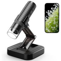
In recent years, advancements in digital imaging technology have allowed for more precise measurements of specimens under a microscope. Digital imaging software can be used to capture images of the specimen and measure its size directly on the computer screen. This eliminates the need for manual estimation and provides more accurate and reproducible results.
In conclusion, there are various measurement techniques available to estimate the size of a specimen under a microscope. These techniques involve the use of calibrated eyepiece reticles, stage micrometers, and digital imaging software. The choice of technique depends on the specific requirements of the study and the available equipment.
2、 Calibration methods for accurate estimation of specimen size
To estimate the size of a specimen under a microscope, calibration methods are used to ensure accurate measurements. These methods involve establishing a relationship between the size of an object in the microscope's field of view and the actual size of the object.
One common calibration method is using a stage micrometer, which is a slide with a known scale etched onto it. By comparing the size of the specimen to the scale on the stage micrometer, the size of the specimen can be estimated. This method requires adjusting the microscope's magnification and focusing on the stage micrometer to obtain accurate measurements.
Another calibration method involves using an eyepiece reticle, which is a small glass disk with a scale etched onto it that fits into the microscope's eyepiece. By aligning the scale on the eyepiece reticle with the specimen and adjusting the microscope's magnification, the size of the specimen can be estimated.
In recent years, digital imaging and software analysis have become increasingly popular for estimating specimen size. With this method, images of the specimen are captured using a digital camera attached to the microscope. The images are then analyzed using software that allows for accurate measurements of the specimen's size. This method offers the advantage of automated measurements and the ability to store and analyze data.
It is important to note that calibration methods should be performed regularly to account for any changes in the microscope's optics or magnification settings. Additionally, it is recommended to use multiple calibration methods and compare the results to ensure accuracy.
In conclusion, calibration methods such as using a stage micrometer, eyepiece reticle, or digital imaging with software analysis are commonly employed to estimate the size of a specimen under a microscope. These methods provide accurate measurements and can be adapted to the latest technological advancements in microscopy.
3、 Factors influencing the estimation of specimen size under a microscope
Estimating the size of a specimen under a microscope is an essential skill in microscopy. It allows scientists and researchers to accurately measure and compare the dimensions of microscopic objects. However, several factors can influence the estimation of specimen size, and understanding these factors is crucial for obtaining reliable measurements.
One of the primary factors influencing the estimation of specimen size is the magnification of the microscope. Magnification refers to the degree to which the image of the specimen is enlarged. Higher magnification allows for more precise measurements, but it also increases the risk of distortion and loss of clarity. Therefore, it is important to select an appropriate magnification level based on the size and characteristics of the specimen.
Another factor to consider is the calibration of the microscope. Microscopes should be calibrated using a stage micrometer or a known reference object to establish a scale for measurements. This calibration ensures that the measurements obtained are accurate and consistent.
The type of microscope used can also affect the estimation of specimen size. Different microscopes, such as compound microscopes or electron microscopes, have varying levels of resolution and magnification capabilities. It is important to choose the appropriate microscope for the specific requirements of the study.
Additionally, the technique used to prepare the specimen can impact its size estimation. Specimens that are improperly mounted or prepared may appear distorted or altered under the microscope, leading to inaccurate size estimations. Proper sample preparation techniques, such as fixation and staining, should be employed to ensure accurate measurements.
Lastly, advancements in technology have introduced new methods for estimating specimen size. Digital imaging and software analysis tools can enhance the accuracy and precision of size estimations. These tools allow for automated measurements and can compensate for potential human errors.
In conclusion, estimating the size of a specimen under a microscope requires careful consideration of various factors. Magnification, calibration, microscope type, specimen preparation, and technological advancements all play a role in obtaining accurate measurements. Staying updated with the latest techniques and tools can further improve the estimation of specimen size and enhance the overall quality of microscopic analysis.

4、 Common errors and challenges in estimating specimen size under a microscope
Estimating the size of a specimen under a microscope is an essential skill in microscopy. However, it is not without its challenges and potential errors. To ensure accurate estimations, it is important to be aware of common errors and adopt the latest techniques.
One common error in estimating specimen size is the lack of calibration. Microscopes often have a built-in calibration scale, but it is crucial to calibrate the microscope for each objective lens used. Failure to do so can lead to inaccurate estimations. Additionally, errors can arise from improper focusing, as the depth of field can affect the perceived size of the specimen.
Another challenge is the distortion caused by the microscope's optics. Optical distortion can occur due to lens aberrations or improper alignment. It is important to regularly check and maintain the microscope's optics to minimize these errors.
The latest point of view in estimating specimen size involves the use of digital imaging and software analysis. Digital microscopes equipped with cameras allow for capturing images of the specimen, which can then be analyzed using specialized software. This approach provides more accurate measurements and eliminates potential human errors.
To estimate the size of a specimen under a microscope accurately, it is recommended to calibrate the microscope, ensure proper focusing, and be aware of potential optical distortions. Embracing digital imaging and software analysis can further enhance accuracy. Regular training and practice in estimation techniques are also crucial to minimize errors and improve proficiency.









There are no comments for this blog.