How To Focus In Microscope ?
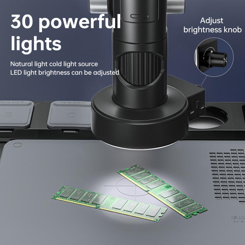
To focus in a microscope, start by placing the slide on the stage and securing it with the stage clips. Turn on the microscope's light source and adjust the intensity if needed. Use the coarse adjustment knob to lower the objective lens close to the slide. Look through the eyepiece and slowly turn the coarse adjustment knob to raise the objective lens until the image comes into view. Once the image is visible, use the fine adjustment knob to sharpen the focus. Adjust the condenser and diaphragm to optimize the lighting and contrast if necessary. Finally, if using a compound microscope with multiple objective lenses, switch to a higher magnification objective and repeat the focusing process.
1、 Adjusting the focus knob for optimal clarity
To focus in a microscope, the first step is to adjust the focus knob for optimal clarity. The focus knob is usually located on the side or underneath the microscope stage. By turning the focus knob, you can move the objective lenses closer or farther away from the specimen, allowing you to achieve a sharp and clear image.
To begin, place your slide on the stage and secure it in place using the stage clips. Start with the lowest magnification objective lens (usually 4x or 10x) and position it over the slide. Look through the eyepiece and use the coarse focus knob to move the objective lens closer to the specimen. As you turn the knob, the image will become blurry at first, but continue to turn until the image starts to come into focus.
Once you have achieved a rough focus, use the fine focus knob to make small adjustments. This will help you fine-tune the focus and bring out more details in the specimen. Slowly turn the fine focus knob in both directions until you find the point where the image appears the sharpest and clearest.
It is important to note that different microscopes may have slightly different mechanisms for focusing. Some microscopes may have separate knobs for coarse and fine focus, while others may have a single focus knob that combines both functions. Additionally, some advanced microscopes may have autofocus capabilities or digital focus adjustments.
In recent years, advancements in microscope technology have led to the development of digital microscopes. These microscopes often have built-in cameras and software that allow for easier focusing and image capture. Some digital microscopes even have autofocus features that automatically adjust the focus for optimal clarity.
In conclusion, adjusting the focus knob for optimal clarity is the key to focusing in a microscope. By using the coarse and fine focus knobs, you can bring the specimen into sharp focus and observe its intricate details. With the advent of digital microscopes, focusing has become even more convenient and precise, offering new possibilities for scientific research and education.

2、 Using the condenser to enhance focus and contrast
To focus in a microscope, one can use the condenser to enhance focus and contrast. The condenser is an important component of the microscope that helps to gather and focus light onto the specimen. By properly adjusting the condenser, one can achieve a clearer and more detailed image.
To begin, start by adjusting the height of the condenser. Raise it as close to the stage as possible without touching the slide. This will ensure that the light is focused properly on the specimen. Next, adjust the iris diaphragm, which controls the amount of light passing through the condenser. Opening the diaphragm will allow more light to pass through, while closing it will reduce the amount of light. Finding the right balance is crucial for achieving optimal focus and contrast.
Once the condenser is properly adjusted, it is important to focus the microscope using the fine focus knob. Start with the lowest magnification objective lens and slowly turn the fine focus knob until the image becomes clear. Then, switch to a higher magnification objective lens and repeat the process. It is important to note that the condenser may need to be readjusted slightly when switching between different magnifications.
In recent years, there have been advancements in microscope technology that have improved focus and contrast. For example, some microscopes now come with adjustable condensers that allow for more precise control of light. Additionally, digital microscopes have become more popular, offering features such as autofocus and image enhancement algorithms that can further improve focus and contrast.
In conclusion, using the condenser to enhance focus and contrast is a crucial step in focusing a microscope. By adjusting the condenser height and iris diaphragm, one can optimize the amount of light reaching the specimen. Additionally, advancements in microscope technology have provided new tools and features to improve focus and contrast, making microscopy even more accessible and accurate.

3、 Employing the fine focus adjustment for precise focusing
To focus in a microscope, one can employ the fine focus adjustment for precise focusing. The fine focus adjustment is a mechanism that allows for small, incremental changes in the focus of the microscope. This adjustment is typically located on the side or underneath the stage of the microscope.
To begin focusing, start with the coarse focus adjustment to bring the specimen into view. Once the specimen is visible, switch to the fine focus adjustment to achieve a sharper and more detailed image. The fine focus adjustment allows for minute changes in the position of the objective lens, resulting in a clearer view of the specimen.
To use the fine focus adjustment effectively, it is important to make small, gradual adjustments. Turning the adjustment knob too quickly or forcefully can result in overshooting the desired focus point and potentially damaging the specimen or the microscope itself. It is recommended to turn the knob slowly and observe the changes in focus as you make adjustments.
Additionally, it is crucial to have proper lighting when using the fine focus adjustment. Insufficient lighting can make it difficult to accurately focus on the specimen. Adjusting the light intensity or angle can help improve visibility and aid in achieving a clear focus.
In recent years, advancements in microscope technology have led to the development of automated focusing systems. These systems use algorithms and sensors to automatically adjust the focus, eliminating the need for manual fine focus adjustments. This technology has greatly improved the efficiency and accuracy of focusing in microscopes, particularly in applications such as digital pathology and high-throughput screening.
In conclusion, employing the fine focus adjustment for precise focusing is a fundamental technique in using a microscope. By making small, gradual adjustments and ensuring proper lighting, one can achieve a clear and detailed view of the specimen. The latest advancements in microscope technology have further enhanced the focusing process, offering automated systems for improved efficiency and accuracy.

4、 Utilizing immersion oil for high-resolution focusing
To focus in a microscope, one can follow a few steps to ensure clear and high-resolution images. One important technique is utilizing immersion oil, which helps improve the resolution and clarity of the specimen being observed.
Firstly, place the slide with the specimen on the microscope stage and secure it in place. Start with the lowest magnification objective lens and bring the specimen into focus using the coarse focus knob. Once the specimen is roughly in focus, switch to the higher magnification objective lens.
To utilize immersion oil, ensure that the lens you are using is designed for oil immersion. These lenses have a higher numerical aperture and are specifically designed to work with oil. Apply a small drop of immersion oil directly onto the slide, directly above the specimen. Then, carefully rotate the nosepiece to bring the oil immersion lens into position above the oil drop.
Next, slowly lower the oil immersion lens until it touches the oil drop. The oil helps to eliminate the air gap between the lens and the slide, reducing light refraction and increasing the numerical aperture. This results in improved resolution and clarity of the specimen.
Finally, use the fine focus knob to bring the specimen into sharp focus. Take care not to apply too much pressure, as this can damage the lens or the slide. Once the specimen is in focus, you can observe and capture high-resolution images.
It is worth noting that while immersion oil is a valuable tool for high-resolution focusing, it is not suitable for all types of microscopy. For example, it is not used in electron microscopy. Additionally, it is important to clean the oil off the lens and slide after use to prevent contamination and damage.
In recent years, advancements in microscopy technology have led to the development of new techniques for high-resolution imaging. For instance, super-resolution microscopy techniques such as stimulated emission depletion (STED) microscopy and structured illumination microscopy (SIM) have allowed researchers to surpass the diffraction limit of light, enabling the visualization of structures at the nanoscale. These techniques have revolutionized the field of microscopy and opened up new possibilities for studying biological processes in unprecedented detail. However, the use of immersion oil remains a fundamental technique for achieving high-resolution focusing in traditional light microscopy.
