How To Identify Cyanobacteria Under Microscope ?
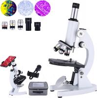
To identify cyanobacteria under a microscope, you can follow these steps:
1. Prepare a microscope slide by placing a small drop of water containing the sample on it.
2. Cover the sample with a coverslip to prevent drying.
3. Start with the lowest magnification objective lens and focus on the sample.
4. Look for cells that appear as small, elongated or spherical structures.
5. Observe the color of the cells, which can range from green to blue-green or even red.
6. Look for the presence of characteristic structures like gas vesicles or heterocysts.
7. Pay attention to the arrangement of cells, whether they are solitary or form colonies or filaments.
8. Take note of any motility, such as gliding or oscillatory movement.
9. Use higher magnification lenses to observe finer details of the cells, such as cell wall structure or presence of chloroplasts.
10. Compare your observations with reference images or descriptions to help identify the cyanobacteria species.
Remember to handle the microscope and slides carefully and adjust the lighting and focus as needed for optimal observation.
1、 Morphological characteristics of cyanobacteria under a microscope
To identify cyanobacteria under a microscope, there are several morphological characteristics that can be observed. These characteristics include cell shape, cell arrangement, presence of sheaths, and the presence of specialized structures such as heterocysts and akinetes.
Cell shape: Cyanobacteria can have various cell shapes, including cylindrical, spherical, filamentous, or colonial. Observing the shape of the cells can provide initial clues for identification.
Cell arrangement: Cyanobacteria can be found as single cells, in chains, or in colonies. The arrangement of cells can vary depending on the species and can be helpful in distinguishing different types of cyanobacteria.
Presence of sheaths: Many cyanobacteria are surrounded by a gelatinous sheath. This sheath can be observed under the microscope as a clear, slimy layer surrounding the cells. The presence and characteristics of the sheath can aid in identification.
Specialized structures: Cyanobacteria may possess specialized structures such as heterocysts and akinetes. Heterocysts are specialized cells involved in nitrogen fixation and can be identified by their larger size and lack of pigmentation. Akinetes are thick-walled resting cells that can withstand harsh environmental conditions and can be identified by their distinct shape and size.
It is important to note that the identification of cyanobacteria under a microscope may require additional techniques such as staining or molecular analysis for accurate species identification. Additionally, the latest point of view in cyanobacteria identification includes the use of molecular techniques such as DNA sequencing to identify specific genetic markers that can differentiate between different species of cyanobacteria. This approach allows for more precise identification and classification of cyanobacteria.

2、 Cellular organization and arrangement of cyanobacteria cells
To identify cyanobacteria under a microscope, there are several key characteristics and techniques that can be employed. The cellular organization and arrangement of cyanobacteria cells can provide important clues for identification.
Firstly, cyanobacteria are prokaryotic organisms, meaning they lack a nucleus and membrane-bound organelles. They typically have a simple cellular structure, consisting of a cell wall and a plasma membrane. Under a microscope, cyanobacteria cells appear as small, elongated or spherical structures.
One important feature to look for is the presence of specialized structures called heterocysts. These are larger, rounded cells that are involved in nitrogen fixation. Heterocysts can be identified by their thicker cell walls and lack of pigmentation compared to other cyanobacteria cells.
Another characteristic to observe is the arrangement of cyanobacteria cells. Some cyanobacteria form colonies or filaments, while others exist as single cells. Filamentous cyanobacteria can be identified by their long, thread-like structures composed of multiple cells attached end-to-end.
In recent years, molecular techniques have become increasingly important in cyanobacteria identification. DNA sequencing can provide valuable information about the genetic makeup of cyanobacteria, aiding in their classification and identification. Additionally, fluorescence microscopy can be used to detect specific pigments or proteins within cyanobacteria cells, providing further insights into their identity.
It is worth noting that the identification of cyanobacteria can be challenging due to their morphological similarities and the existence of cryptic species. Therefore, a combination of traditional microscopy techniques and molecular methods is often necessary for accurate identification.

3、 Identification of cyanobacteria through pigmentation and coloration
To identify cyanobacteria under a microscope, there are several key characteristics to look for. One of the most important factors is pigmentation and coloration. Cyanobacteria are known for their ability to produce various pigments, which can give them distinct colors. These pigments include chlorophyll a (green), phycocyanin (blue), phycoerythrin (red), and carotenoids (yellow, orange, or red).
When observing cyanobacteria under a microscope, it is essential to use appropriate staining techniques to enhance the visibility of these pigments. For example, using a technique called the Ziehl-Neelsen stain can help differentiate between different types of cyanobacteria based on their pigmentation. This stain specifically targets the cell walls of cyanobacteria, making them more visible.
Additionally, the latest point of view in cyanobacteria identification involves the use of molecular techniques. DNA sequencing and genetic analysis can provide more accurate identification of cyanobacteria species. This approach allows for the detection of specific genetic markers that are unique to different cyanobacteria species, enabling more precise identification.
It is worth noting that the identification of cyanobacteria can be challenging due to their morphological similarities. Therefore, a combination of pigmentation analysis, staining techniques, and molecular methods is often necessary for accurate identification. Furthermore, it is crucial to consult taxonomic experts or reference databases to confirm the identification of cyanobacteria species, as new discoveries and taxonomic revisions are continuously being made in this field.

4、 Recognizing cyanobacterial colonies and growth patterns
To identify cyanobacteria under a microscope, there are several key characteristics and techniques to consider. Recognizing cyanobacterial colonies and growth patterns is crucial in distinguishing them from other types of bacteria. Here is a step-by-step guide on how to identify cyanobacteria under a microscope:
1. Sample collection: Collect a water or soil sample from the desired location. Cyanobacteria are commonly found in freshwater bodies, such as lakes, ponds, and rivers.
2. Slide preparation: Take a small amount of the collected sample and place it on a microscope slide. Add a drop of water to create a thin film and cover it with a coverslip.
3. Microscope observation: Use a compound microscope with a magnification of at least 400x. Observe the slide under brightfield illumination.
4. Morphological features: Look for specific morphological features of cyanobacteria, such as filamentous or colonial growth patterns. Cyanobacteria often form long chains or clusters of cells.
5. Pigmentation: Observe the coloration of the cells. Cyanobacteria can exhibit various pigments, including green, blue-green, brown, or red, due to the presence of chlorophyll and other accessory pigments.
6. Cell structure: Pay attention to the cell structure. Cyanobacteria have prokaryotic cells with distinct cell walls and lack a nucleus. They may also possess specialized structures like heterocysts or akinetes.
7. Use of stains: If necessary, use specific stains like Lugol's iodine or methylene blue to enhance the visibility of cyanobacteria cells and structures.
It is important to note that the identification of cyanobacteria under a microscope can be challenging due to their morphological similarities with other microorganisms. Therefore, it is recommended to consult taxonomic keys, specialized literature, or seek expert assistance to confirm the identification. Additionally, advancements in molecular techniques, such as DNA sequencing, can provide more accurate identification of cyanobacteria species.


