How To Make An Endoscope ?
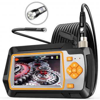
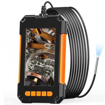
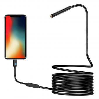
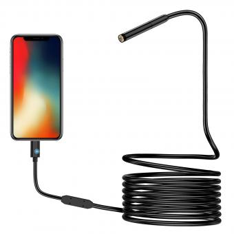
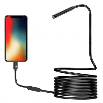
Making an endoscope requires specialized knowledge and equipment. Endoscopes are medical devices used to examine the inside of the body, and they consist of a long, thin tube with a camera and light source at the end. The tube is inserted into the body through a natural opening or a small incision.
The process of making an endoscope involves designing and manufacturing the tube, camera, and light source, as well as assembling and testing the device. The tube must be flexible enough to navigate through the body, but also strong enough to withstand repeated use. The camera and light source must be high-quality and able to provide clear images.
Endoscopes are typically made by medical device companies that specialize in this type of equipment. The process involves a team of engineers, designers, and technicians who work together to create a safe and effective device. The manufacturing process is highly regulated to ensure that the endoscope meets strict quality and safety standards.
1、 Optics and lens assembly
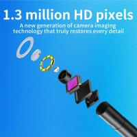
How to make an endoscope involves several steps, but one of the most critical components is the optics and lens assembly. This assembly is responsible for capturing high-quality images of the internal body parts and transmitting them to the viewing screen.
To make an endoscope, the optics and lens assembly must be designed and manufactured with precision. The assembly typically consists of a series of lenses, mirrors, and fiber optic cables that work together to transmit light and images.
The latest point of view in endoscope technology is the use of digital imaging systems. These systems use advanced sensors and processors to capture and process high-resolution images in real-time. This technology has revolutionized the field of endoscopy, allowing for more accurate diagnoses and better patient outcomes.
To manufacture the optics and lens assembly, specialized equipment and techniques are required. The lenses must be ground and polished to exact specifications, and the fiber optic cables must be carefully aligned to ensure optimal light transmission.
Once the assembly is complete, it must be tested and calibrated to ensure that it meets the required standards for image quality and performance. This involves using specialized testing equipment and procedures to evaluate the assembly's resolution, contrast, and distortion.
In conclusion, making an endoscope requires a high level of expertise and precision, particularly when it comes to the optics and lens assembly. With the latest advancements in digital imaging technology, endoscopes are becoming more accurate and effective than ever before, improving patient outcomes and advancing the field of medicine.
2、 Light source and illumination system

Light source and illumination system are crucial components in the construction of an endoscope. The light source provides the necessary illumination to visualize the internal organs or cavities, while the illumination system ensures that the light is directed to the area of interest.
To make an endoscope, the first step is to select the appropriate light source. LED lights are commonly used in modern endoscopes due to their low power consumption, long lifespan, and high brightness. The LED light is then connected to a fiber optic cable, which transmits the light to the endoscope's distal end.
The illumination system consists of a series of lenses and mirrors that direct the light to the area of interest. The lenses and mirrors are carefully positioned to ensure that the light is evenly distributed and focused on the target area. The illumination system also includes a light guide that directs the light from the fiber optic cable to the lenses and mirrors.
In recent years, there have been significant advancements in endoscope technology, including the development of wireless endoscopes that do not require a physical connection to the light source. These endoscopes use miniature LED lights and micro-optics to provide illumination, making them more compact and portable than traditional endoscopes.
In conclusion, the light source and illumination system are essential components in the construction of an endoscope. The latest advancements in endoscope technology have led to the development of wireless endoscopes that are more compact and portable than traditional endoscopes.
3、 Camera and image sensor technology
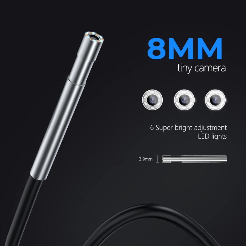
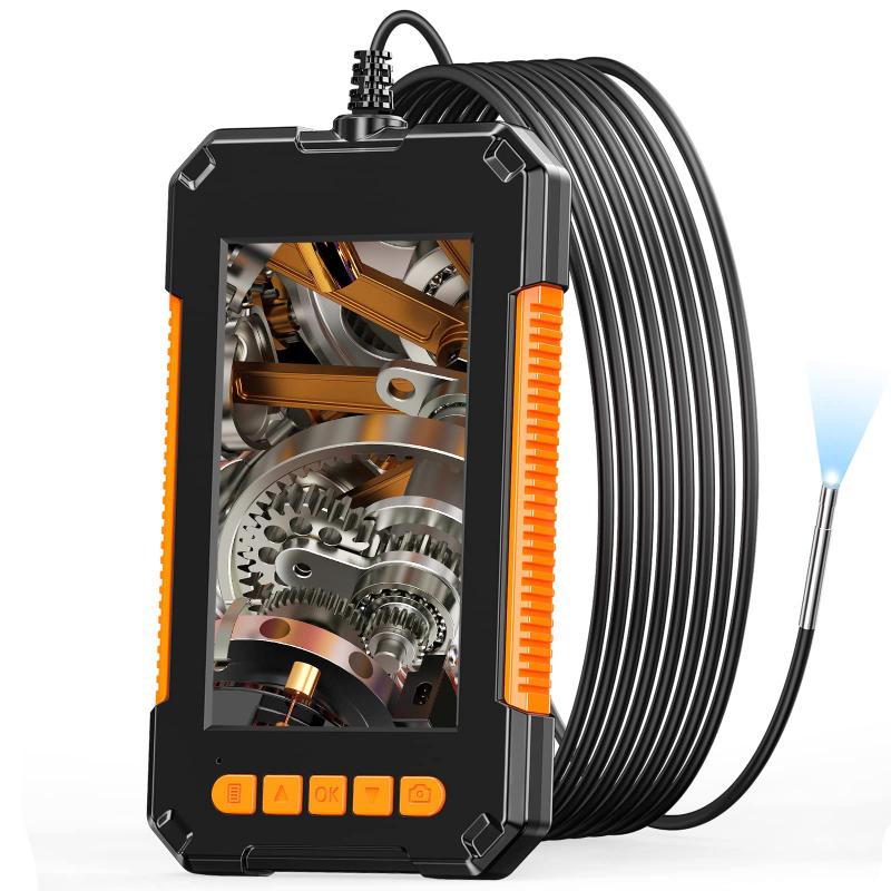
How to make an endoscope involves the use of camera and image sensor technology. An endoscope is a medical device used to examine the inside of the body, particularly the digestive tract. It consists of a long, thin, flexible tube with a camera and light source at the end. The camera captures images of the inside of the body, which are transmitted to a monitor for viewing by the doctor.
To make an endoscope, the first step is to select the appropriate camera and image sensor technology. The camera must be small enough to fit inside the tube and capable of capturing high-quality images. The image sensor must be sensitive enough to capture clear images in low-light conditions.
Once the camera and image sensor technology have been selected, the next step is to design the endoscope tube. The tube must be flexible enough to navigate through the body's curves and narrow passages, yet strong enough to withstand repeated use.
The camera and light source are then attached to the end of the tube. The camera captures images of the inside of the body, while the light source illuminates the area being examined.
The latest point of view in endoscope technology is the use of wireless endoscopes. These devices use wireless technology to transmit images from the camera to a monitor, eliminating the need for a physical cable. This makes the endoscope more flexible and easier to use.
In conclusion, making an endoscope involves the use of camera and image sensor technology. The latest point of view in endoscope technology is the use of wireless endoscopes, which offer greater flexibility and ease of use.
4、 Control and manipulation mechanisms
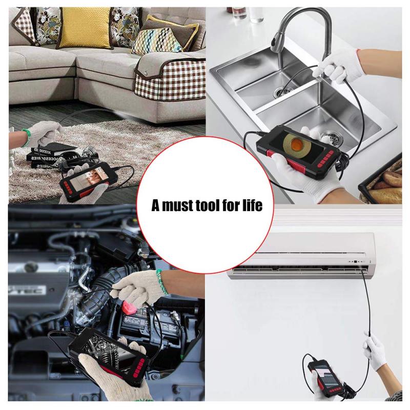
How to make an endoscope:
An endoscope is a medical device used to examine the inside of the body. It consists of a long, thin tube with a camera and light source at the end. The tube is inserted into the body through a natural opening or a small incision. The camera sends images to a monitor, allowing doctors to see inside the body.
To make an endoscope, the following steps are typically involved:
1. Design: The first step is to design the endoscope. This involves determining the size, shape, and features of the device. The design must be precise to ensure that the endoscope can reach the desired area of the body and provide clear images.
2. Materials: The endoscope is typically made of a flexible or rigid tube, a camera, a light source, and control and manipulation mechanisms. The materials used must be durable, lightweight, and safe for use in the body.
3. Assembly: The components of the endoscope are assembled by skilled technicians. The camera and light source are connected to the tube, and the control and manipulation mechanisms are added.
4. Testing: The endoscope is tested to ensure that it functions properly. This involves checking the camera and light source, as well as the control and manipulation mechanisms.
Control and manipulation mechanisms:
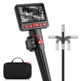
The control and manipulation mechanisms of an endoscope allow doctors to move the device inside the body and adjust the camera angle. These mechanisms typically include knobs or buttons that are located on the handle of the endoscope. By turning or pressing these controls, doctors can move the endoscope up, down, left, or right, and rotate the camera to get a better view.
The latest point of view:
Recent advancements in endoscope technology have led to the development of more advanced control and manipulation mechanisms. For example, some endoscopes now use robotic arms to move the device inside the body. These arms are controlled by a computer, allowing for more precise movements and greater flexibility. Additionally, some endoscopes now use virtual reality technology to provide doctors with a more immersive view of the inside of the body. This technology allows doctors to see the body in 3D, which can help with diagnosis and treatment planning.


