How To Observe A Specimen Under A Microscope ?
To observe a specimen under a microscope, first, prepare the specimen by placing it on a glass slide. Add a drop of water or a suitable mounting medium to keep the specimen in place. Carefully cover the specimen with a coverslip to avoid any air bubbles. Adjust the focus and lighting of the microscope to obtain a clear image. Start with the lowest magnification objective lens and gradually increase the magnification as needed. Move the slide around using the mechanical stage knobs to explore different areas of the specimen. Use the focus knobs to bring the specimen into sharp focus. Observe the specimen carefully, noting its structure, shape, and any other relevant details. You can also use additional techniques like staining or using different microscopy techniques to enhance the visibility of specific features in the specimen.
1、 Preparation of the specimen for microscopy
Preparation of the specimen for microscopy is a crucial step in observing a specimen under a microscope. It ensures that the specimen is properly prepared and mounted on a slide, allowing for clear and accurate observations. Here is a step-by-step guide on how to prepare a specimen for microscopy:
1. Collection: Start by collecting the specimen you wish to observe. This can be anything from a plant leaf to a microorganism. Ensure that the specimen is fresh and intact for the best results.
2. Fixation: Fixation is the process of preserving the specimen's structure and preventing decay. It involves immersing the specimen in a fixative solution, such as formaldehyde or ethanol. The choice of fixative depends on the type of specimen and the desired observations.
3. Dehydration: Dehydration removes water from the specimen, allowing it to be properly mounted on a slide. This is typically done by gradually replacing the water in the specimen with an alcohol series, starting with lower concentrations and increasing to higher concentrations.
4. Clearing: Clearing is the process of making the specimen transparent. It involves using a clearing agent, such as xylene or cedarwood oil, to replace the alcohol in the specimen. This step is important for enhancing the visibility of the specimen under the microscope.
5. Mounting: Once the specimen is dehydrated and cleared, it can be mounted on a slide. Place a small drop of mounting medium, such as glycerin or a mounting gel, on a clean slide. Carefully transfer the specimen onto the slide, ensuring it is properly positioned.
6. Coverslipping: To protect the specimen and prevent it from drying out, place a coverslip over the mounted specimen. Gently lower the coverslip onto the slide, avoiding any air bubbles. Press down lightly to ensure a secure seal.
7. Labeling: Finally, label the slide with relevant information, such as the specimen's name, date, and any additional details. This is important for proper documentation and future reference.
It is worth noting that advancements in microscopy techniques, such as confocal microscopy and super-resolution microscopy, have revolutionized the field. These techniques allow for even more detailed and precise observations of specimens, providing researchers with a deeper understanding of biological structures and processes.
2、 Adjusting the microscope settings for optimal observation
Observing a specimen under a microscope requires careful preparation and adjustment of the microscope settings to ensure optimal observation. Here is a step-by-step guide on how to observe a specimen under a microscope:
1. Prepare the specimen: Start by preparing the specimen you want to observe. This may involve fixing, staining, or mounting the specimen on a slide, depending on the type of specimen.
2. Place the slide on the stage: Gently place the prepared slide on the stage of the microscope. Ensure that the specimen is centered and secured in place.
3. Start with low magnification: Begin by using the lowest magnification objective lens. This will provide a wider field of view and make it easier to locate the specimen.
4. Adjust the lighting: Adjust the light intensity to ensure proper illumination of the specimen. Use the condenser to control the amount of light passing through the specimen.
5. Focus on the specimen: Use the coarse adjustment knob to bring the specimen into rough focus. Then, use the fine adjustment knob to achieve a clear and sharp image of the specimen.
6. Increase magnification: Once the specimen is in focus, gradually increase the magnification by switching to higher objective lenses. Refocus the specimen at each magnification level.
7. Adjust the diaphragm: Depending on the type of specimen and the desired contrast, adjust the diaphragm to control the amount of light entering the microscope. This can enhance the visibility of certain structures within the specimen.
8. Take notes or capture images: If necessary, take notes or capture images of the observed specimen for further analysis or documentation.
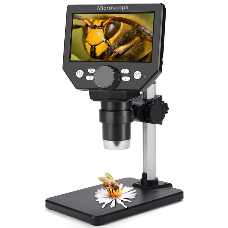
It is important to note that the latest advancements in microscopy technology, such as digital microscopes and confocal microscopy, have revolutionized the way we observe specimens. These technologies offer higher resolution, 3D imaging, and the ability to observe live specimens in real-time. However, the basic principles of adjusting microscope settings for optimal observation remain the same.

3、 Focusing and centering the specimen on the microscope slide
Observing a specimen under a microscope is a fundamental skill in the field of microscopy. It allows scientists, researchers, and students to explore the intricate details of various biological and non-biological samples. One of the initial steps in this process is focusing and centering the specimen on the microscope slide.
To begin, ensure that the microscope is properly set up and adjusted for optimal viewing. Place the prepared slide on the stage and secure it with the stage clips. Start with the lowest magnification objective lens and adjust the coarse focus knob to bring the specimen into view. This initial adjustment will provide a rough focus of the specimen.
Next, fine-tune the focus by using the fine focus knob. Slowly turn the knob in both directions until the specimen becomes clear and sharp. It is important to make small adjustments to avoid overshooting the focus point. If the specimen is not in focus, repeat the process of adjusting the coarse and fine focus knobs until a clear image is obtained.
Once the specimen is in focus, center it on the microscope slide. Use the mechanical stage controls to move the slide horizontally and vertically until the specimen is positioned in the center of the field of view. This ensures that the entire specimen can be observed without having to constantly readjust the slide.
In recent years, advancements in microscopy technology have introduced automated focusing and centering systems. These systems utilize computer algorithms and motorized stages to quickly and accurately focus and center the specimen. They provide a more efficient and precise way of observing specimens, especially when dealing with large datasets or high-throughput experiments.
In conclusion, focusing and centering the specimen on the microscope slide is a crucial step in observing a specimen under a microscope. It allows for clear visualization and ensures that the entire specimen is within the field of view. While manual adjustments are still widely used, automated systems offer enhanced efficiency and accuracy in the observation process.

4、 Using different magnifications to observe different details of the specimen
Observing a specimen under a microscope is a fundamental technique in the field of biology and other scientific disciplines. It allows scientists to study the intricate details of cells, tissues, and other microscopic structures. Here is a step-by-step guide on how to observe a specimen under a microscope:
1. Prepare the specimen: Start by obtaining a small sample of the specimen you wish to observe. This can be a thin slice of tissue, a drop of liquid containing microorganisms, or any other suitable sample. Ensure that the specimen is properly prepared, such as fixing and staining, to enhance visibility and contrast.
2. Set up the microscope: Place the specimen slide on the stage of the microscope and secure it with the stage clips. Adjust the focus knobs to bring the specimen into rough focus.
3. Start with low magnification: Begin observing the specimen using the lowest magnification objective lens (usually 4x or 10x). This will provide an overview of the specimen and help you locate areas of interest.
4. Increase magnification: Once you have identified an area of interest, switch to a higher magnification objective lens (such as 40x or 100x). This will allow you to observe finer details of the specimen.
5. Adjust the focus: Use the fine focus knob to bring the specimen into sharp focus. This may require slight adjustments as you switch between different magnifications.
6. Observe and record: Take your time to carefully observe the specimen under the microscope. Pay attention to the structure, shape, and arrangement of cells or other components. Make notes or take photographs to document your observations.
Using different magnifications to observe different details of the specimen is crucial. Lower magnifications provide a broader view, allowing you to observe the overall structure and organization of the specimen. Higher magnifications, on the other hand, enable you to examine finer details such as individual cells, organelles, or cellular processes.
It is important to note that the latest advancements in microscopy technology have revolutionized the field of observation. Techniques such as confocal microscopy, electron microscopy, and super-resolution microscopy have pushed the boundaries of what can be observed under a microscope. These techniques offer enhanced resolution, improved contrast, and the ability to observe dynamic processes in real-time.
In conclusion, observing a specimen under a microscope involves preparing the specimen, setting up the microscope, starting with low magnification, increasing magnification to observe finer details, adjusting focus, and carefully observing and documenting your findings. The use of different magnifications is essential to explore various aspects of the specimen, and the latest advancements in microscopy technology continue to expand our understanding of the microscopic world.
