How To See Cells In A Microscope?
To see cells in a microscope, you need to prepare a sample of the cells and place it on a microscope slide. The sample can be obtained from a tissue culture, blood, or other biological material. The cells are then stained with a dye that will make them visible under the microscope. The most commonly used dyes are hematoxylin and eosin (H&E), which stain the nucleus and cytoplasm of the cells, respectively.
Once the sample is prepared and stained, it is placed on the stage of the microscope and viewed through the eyepiece. The microscope has several lenses that magnify the image of the cells, allowing you to see them in detail. You can adjust the focus and the magnification of the microscope to get a clearer view of the cells.
It is important to handle the microscope and the sample carefully to avoid damaging the cells or the microscope. You should also clean the microscope after use to prevent contamination of future samples.
1、 Microscope types and parts

How to see cells in a microscope:
To see cells in a microscope, you will need to follow these steps:
1. Prepare a sample: You can prepare a sample by taking a small piece of tissue or a drop of liquid containing cells and placing it on a microscope slide.
2. Stain the sample: Staining the sample will help to highlight the cells and make them easier to see. There are different types of stains that can be used depending on the type of cells you are looking at.
3. Place the slide on the microscope: Once the sample is prepared and stained, place the slide on the microscope stage and secure it in place.
4. Adjust the focus: Use the coarse and fine focus knobs to adjust the focus until the cells come into view.
5. Observe the cells: Once the cells are in focus, you can observe them and take measurements or make notes about their characteristics.
Microscope types and parts:
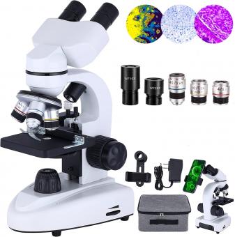
There are several types of microscopes, including compound microscopes, stereo microscopes, and electron microscopes. Compound microscopes are the most commonly used type and are used to view small specimens such as cells. Stereo microscopes are used for larger specimens and provide a 3D view. Electron microscopes use a beam of electrons to view specimens at a much higher magnification than other types of microscopes.
The main parts of a microscope include the eyepiece, objective lenses, stage, focus knobs, and light source. The eyepiece is where you look through to view the specimen. The objective lenses are located on a rotating nosepiece and provide different levels of magnification. The stage is where the specimen is placed and can be moved up and down or side to side to adjust the view. The focus knobs are used to adjust the focus of the specimen. The light source provides illumination for the specimen.
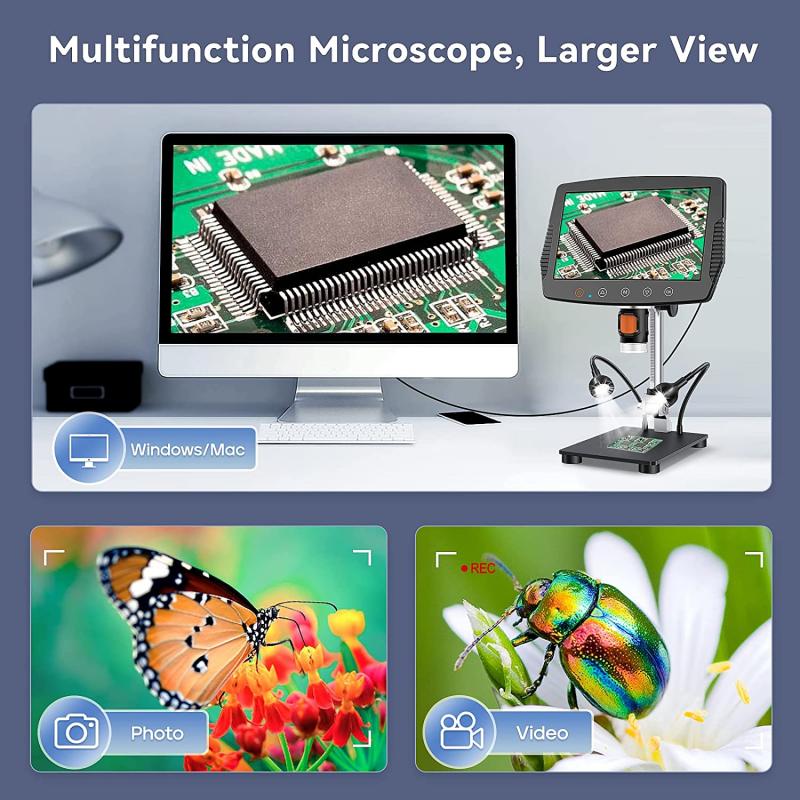
In recent years, advancements in technology have led to the development of new types of microscopes, such as super-resolution microscopes, which can provide even higher levels of magnification and resolution. Additionally, digital microscopes have become more popular, allowing for images and videos of specimens to be captured and shared electronically.
2、 Sample preparation techniques

How to see cells in a microscope:
To see cells in a microscope, sample preparation techniques are crucial. The following steps can be taken to prepare a sample for viewing under a microscope:
1. Collect the sample: The sample can be collected from a variety of sources, such as tissue, blood, or bacteria.
2. Fixation: The sample needs to be fixed to preserve its structure. This can be done by using chemicals such as formaldehyde or glutaraldehyde.
3. Dehydration: The sample needs to be dehydrated to remove any water content. This can be done by using a series of alcohol solutions with increasing concentrations.
4. Embedding: The sample needs to be embedded in a medium that will allow it to be sliced thinly for viewing under a microscope. This can be done by using paraffin wax or resin.
5. Sectioning: The embedded sample needs to be sliced thinly using a microtome.
6. Staining: The sample needs to be stained to enhance its visibility under a microscope. This can be done by using dyes such as hematoxylin and eosin.
7. Mounting: The sample needs to be mounted on a slide and covered with a coverslip.
The latest point of view in sample preparation techniques is the use of 3D printing technology to create custom molds for embedding samples. This allows for more precise and consistent sample preparation, which can improve the accuracy of microscopy analysis. Additionally, new staining techniques are being developed that use fluorescent dyes to label specific structures within cells, allowing for more detailed imaging.
3、 Adjusting focus and magnification

To see cells in a microscope, there are a few steps that need to be followed. The first step is to prepare a sample of the cells that you want to observe. This can be done by taking a small piece of tissue or a drop of liquid containing the cells and placing it on a microscope slide. The sample can then be covered with a cover slip to protect it and keep it in place.
Once the sample is prepared, the microscope can be set up. The microscope should be placed on a stable surface and the eyepiece and objective lenses should be cleaned to ensure clear viewing. The sample slide can then be placed on the stage of the microscope and secured in place.
The next step is to adjust the focus and magnification of the microscope. This can be done by using the coarse and fine focus knobs to bring the sample into focus. The magnification can be adjusted by rotating the objective lenses to the desired level.
It is important to note that different types of microscopes may have different methods for adjusting focus and magnification. For example, some microscopes may have digital controls for adjusting these settings.
In recent years, advances in technology have led to the development of new types of microscopes, such as confocal microscopes and super-resolution microscopes. These types of microscopes offer even greater levels of detail and precision in observing cells and other microscopic structures.
Overall, seeing cells in a microscope requires careful preparation of the sample and proper adjustment of the microscope's focus and magnification settings. With the right techniques and equipment, researchers can gain valuable insights into the structure and function of cells and other microscopic structures.
4、 Illumination and contrast methods

To see cells in a microscope, there are two main factors to consider: illumination and contrast methods.
Illumination refers to the source of light used to illuminate the sample. The most common type of illumination is brightfield illumination, where light passes through the sample and is absorbed or scattered by the cells. This method is useful for observing the overall shape and size of cells, but it can be difficult to distinguish between different types of cells or structures within cells.
Contrast methods are used to enhance the visibility of cells and their structures. One common method is staining, where cells are treated with dyes that bind to specific structures, such as the nucleus or cell membrane. This allows for better visualization of the cells and their components. Another method is phase contrast microscopy, which uses differences in refractive index to create contrast between different parts of the cell. This method is particularly useful for observing live cells, as it does not require staining or fixation.
In recent years, advances in microscopy technology have allowed for even greater visualization of cells. Super-resolution microscopy techniques, such as structured illumination microscopy and stochastic optical reconstruction microscopy, allow for imaging at the nanoscale level. This has led to new insights into the structure and function of cells, and has the potential to revolutionize our understanding of biology and disease.


