How To See Dna Under A Microscope ?
It is not possible to see DNA under a regular light microscope as DNA molecules are too small to be visible with this type of microscope. However, scientists can use specialized techniques such as electron microscopy or fluorescence microscopy to visualize DNA. In electron microscopy, a beam of electrons is used instead of light to create an image of the DNA molecule. In fluorescence microscopy, fluorescent dyes are used to label the DNA, which can then be visualized under a microscope equipped with a fluorescent filter. These techniques allow scientists to study the structure and behavior of DNA in detail.
1、 DNA Extraction
How to see DNA under a microscope:
To see DNA under a microscope, you first need to extract it from the cells. DNA extraction involves breaking open the cells and separating the DNA from other cellular components. There are many different methods for DNA extraction, but most involve a combination of physical and chemical techniques.
Once you have extracted the DNA, you can visualize it under a microscope using a technique called fluorescence microscopy. This involves staining the DNA with a fluorescent dye that binds specifically to DNA molecules. When you shine a specific wavelength of light on the stained DNA, it emits a bright fluorescent signal that can be visualized using a microscope.
Recent advances in microscopy technology have made it possible to visualize DNA at higher resolutions than ever before. For example, super-resolution microscopy techniques such as STED and PALM can achieve resolutions of a few nanometers, allowing researchers to see individual DNA molecules and even specific regions of the genome.
Overall, visualizing DNA under a microscope is an important tool for understanding the structure and function of the genome. It can help researchers study the organization of DNA within cells, track the movement of DNA during cell division, and investigate the interactions between DNA and other cellular components.
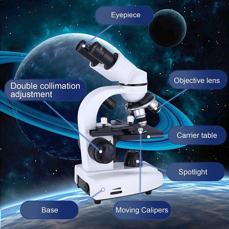
2、 DNA Purification
How to see DNA under a microscope:
To see DNA under a microscope, it must first be extracted and purified from the sample. This can be done using various methods such as phenol-chloroform extraction, column-based purification, or magnetic bead-based purification. Once the DNA is purified, it can be visualized under a microscope using various staining techniques.
One common staining technique is the use of ethidium bromide, which intercalates between the base pairs of DNA and fluoresces under UV light. Another technique is the use of DAPI (4',6-diamidino-2-phenylindole), which binds to the minor groove of DNA and also fluoresces under UV light.
It is important to note that while DNA can be visualized under a microscope, it is not possible to see the actual sequence of the DNA. Instead, the visualization provides information on the size and quantity of the DNA present in the sample.
Latest point of view:
Advancements in technology have allowed for the visualization of DNA at higher resolutions and with greater accuracy. For example, super-resolution microscopy techniques such as STORM (stochastic optical reconstruction microscopy) and PALM (photoactivated localization microscopy) have enabled the visualization of DNA at the nanoscale level.
Additionally, the use of fluorescent in situ hybridization (FISH) allows for the visualization of specific DNA sequences within a sample. This technique involves the use of fluorescently labeled probes that bind to complementary DNA sequences, allowing for the visualization of specific regions of the genome.
Overall, the visualization of DNA under a microscope continues to be an important tool in molecular biology research, providing valuable insights into the structure and function of the genome.
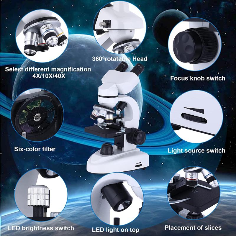
3、 DNA Quantification
How to see DNA under a microscope:
To see DNA under a microscope, the first step is to extract the DNA from the sample. This can be done using various methods, such as using a commercial DNA extraction kit or a homemade method using household items. Once the DNA is extracted, it can be visualized under a microscope using various staining techniques.
One common staining technique is using ethidium bromide, which intercalates between the DNA base pairs and fluoresces under UV light. Another technique is using DAPI (4',6-diamidino-2-phenylindole), which binds to the minor groove of DNA and also fluoresces under UV light.
It is important to note that while DNA can be visualized under a microscope, it is not possible to see the actual sequence of the DNA. Instead, the visualization provides information on the quantity and quality of the DNA sample.
DNA Quantification:
DNA quantification is the process of determining the concentration of DNA in a sample. This is important for various applications, such as PCR (polymerase chain reaction) and sequencing, where a specific amount of DNA is required for optimal results.
There are various methods for DNA quantification, such as using spectrophotometry, fluorometry, and gel electrophoresis. Spectrophotometry measures the absorbance of DNA at a specific wavelength, while fluorometry measures the fluorescence of DNA-binding dyes. Gel electrophoresis separates DNA fragments based on size and can be used to estimate the concentration of DNA in a sample.
The latest point of view on DNA quantification is the use of digital PCR (dPCR), which provides absolute quantification of DNA molecules in a sample. This method is more precise and sensitive than traditional PCR and can be used for various applications, such as detecting rare mutations and measuring gene expression levels.
In conclusion, visualizing DNA under a microscope and quantifying DNA concentration are important techniques in molecular biology. The latest advancements in technology, such as dPCR, have improved the accuracy and sensitivity of DNA quantification.
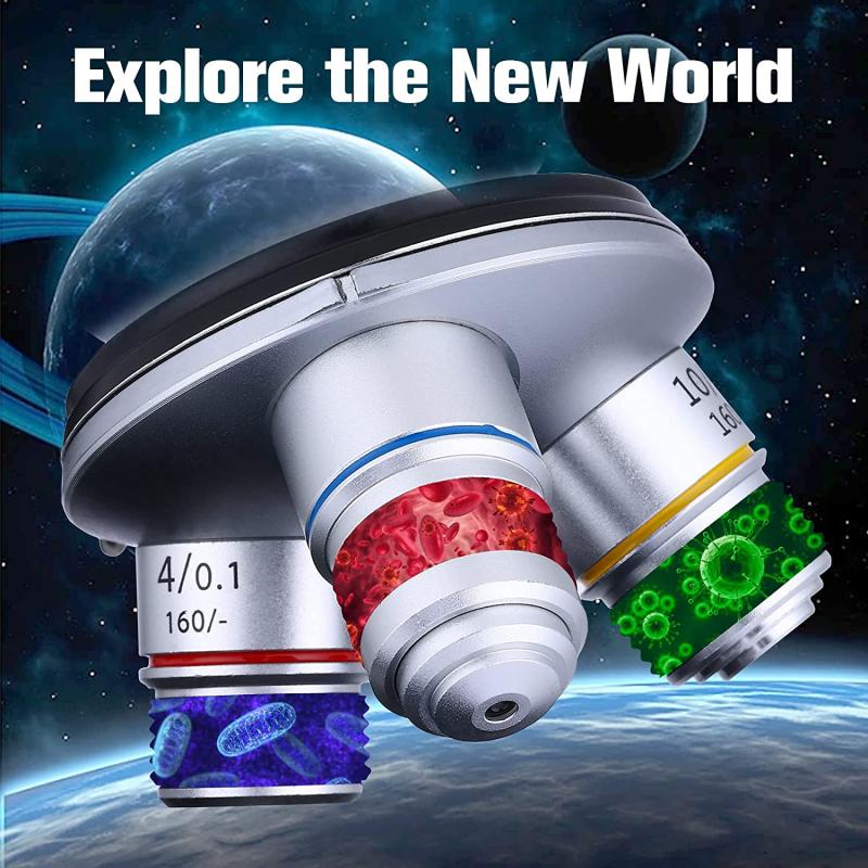
4、 DNA Amplification
How to see DNA under a microscope:
To see DNA under a microscope, it needs to be extracted from cells and purified. Once purified, the DNA can be stained with a fluorescent dye, such as DAPI, which binds to the DNA and makes it visible under a microscope. The DNA can then be viewed using a fluorescence microscope, which uses a specific wavelength of light to excite the fluorescent dye and produce an image of the DNA.
However, it is important to note that DNA is extremely small and difficult to see under a standard light microscope. Therefore, specialized techniques such as electron microscopy may be required to visualize DNA at higher magnifications.
DNA Amplification:
DNA amplification is the process of making multiple copies of a specific DNA sequence. This technique is commonly used in research and diagnostic applications, such as PCR (polymerase chain reaction) and DNA sequencing.
PCR is a widely used DNA amplification technique that allows for the rapid and efficient amplification of a specific DNA sequence. This technique involves the use of a DNA polymerase enzyme, which synthesizes new DNA strands using a template DNA sequence and specific primers that bind to the target sequence.
The latest point of view on DNA amplification is the development of new techniques, such as digital PCR and CRISPR-Cas9, which offer improved sensitivity and specificity for DNA amplification and detection. These techniques have the potential to revolutionize the field of molecular biology and have important applications in areas such as disease diagnosis and personalized medicine.
