How To See Germs Under A Microscope ?
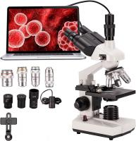
To see germs under a microscope, you will need a compound microscope with a magnification of at least 400x. First, prepare a sample of the germs you want to observe. This can be done by swabbing a surface or collecting a sample of bodily fluids. Next, place the sample on a microscope slide and add a drop of water or stain to enhance visibility. Then, cover the sample with a cover slip to prevent it from drying out and to keep it in place.
Once the sample is prepared, place it on the microscope stage and adjust the focus until the germs come into view. It may take some practice to get the focus just right, so be patient and make small adjustments until the germs are clear and in focus. Finally, observe the germs and take note of their size, shape, and any other characteristics that may be of interest. Remember to properly clean and sterilize the microscope and any tools used to prepare the sample to prevent contamination.
1、 Microbial morphology and structure
How to see germs under a microscope:
To see germs under a microscope, you will need a compound microscope with a high magnification power. First, prepare a sample of the germ you want to observe. This can be done by taking a swab of the area where the germ is suspected to be present, such as a surface or a wound. The swab can then be placed on a microscope slide and stained with a suitable stain, such as Gram stain or acid-fast stain, to make the germs visible.
Once the slide is prepared, place it under the microscope and adjust the focus until the germs come into view. Depending on the type of germ, you may be able to observe its morphology and structure, such as its shape, size, and any unique features it may have, such as flagella or capsules.
It is important to note that not all germs can be seen under a microscope, as some are too small to be resolved with light microscopy. In recent years, advances in electron microscopy have allowed for the visualization of even smaller microbes, such as viruses and prions.
In addition, the field of microbial morphology and structure is constantly evolving, with new techniques and technologies being developed to better understand the complex structures and functions of microbes. For example, super-resolution microscopy techniques, such as stimulated emission depletion (STED) microscopy and structured illumination microscopy (SIM), have allowed for the visualization of structures at the nanoscale level.
Overall, seeing germs under a microscope can provide valuable insights into their morphology and structure, which can help in the diagnosis and treatment of infectious diseases.

2、 Bacterial classification and identification
How to see germs under a microscope:
To see germs under a microscope, you will need a compound microscope with a high magnification power. First, prepare a sample of the germ you want to observe. This can be done by taking a swab of the area where the germ is suspected to be present, such as a surface or a wound. The swab should then be placed on a slide and stained with a suitable stain, such as Gram stain or acid-fast stain, depending on the type of germ being observed.
Once the slide is prepared, it can be placed under the microscope and observed at high magnification. Bacteria can be classified and identified based on their shape, size, and staining characteristics. For example, Gram-positive bacteria will appear purple under the microscope, while Gram-negative bacteria will appear pink.
Bacterial classification and identification:
Bacterial classification and identification have evolved over time with the development of new techniques and technologies. Traditional methods of bacterial identification relied on phenotypic characteristics such as morphology, staining, and biochemical tests. However, these methods have limitations and may not be able to identify all bacterial species.
With the advent of molecular techniques such as polymerase chain reaction (PCR) and DNA sequencing, bacterial identification has become more accurate and reliable. These methods can identify bacteria based on their genetic makeup, allowing for more precise classification and identification.
In recent years, there has also been a growing interest in the study of the human microbiome, which refers to the collection of microorganisms that live in and on the human body. Advances in bacterial classification and identification have allowed for a better understanding of the role of these microorganisms in human health and disease.

3、 Viral replication and pathogenesis
How to see germs under a microscope:
To see germs under a microscope, you will need a compound microscope with a high magnification power. First, prepare a sample of the germ you want to observe. This can be done by taking a swab of the area where the germ is suspected to be present, such as a surface or a bodily fluid. The swab can then be placed on a slide and stained with a special dye that will make the germ visible under the microscope.
Once the slide is prepared, place it on the microscope stage and adjust the focus until the germ comes into view. Depending on the type of germ, it may appear as a small, round shape or a more complex structure.
Viral replication and pathogenesis:
Viral replication is the process by which a virus reproduces itself within a host cell. This process involves the virus attaching to a host cell, entering the cell, and using the cell's machinery to replicate its genetic material and produce new virus particles. The new virus particles can then infect other cells and continue the cycle of replication.
Pathogenesis refers to the process by which a virus causes disease in a host organism. This can occur through a variety of mechanisms, including direct damage to cells, activation of the immune system, and interference with normal cellular processes.
Recent research has shed light on the complex interactions between viruses and host cells, including the role of host factors in determining the outcome of infection. Understanding these processes is critical for the development of effective treatments and vaccines for viral diseases.

4、 Fungal growth and reproduction
How to see germs under a microscope:
To see germs under a microscope, you will need a compound microscope with a high magnification power. First, prepare a sample of the germ you want to observe. This can be done by swabbing a surface or collecting a sample of bodily fluid. Place the sample on a microscope slide and add a drop of water or stain to enhance visibility. Then, place the slide under the microscope and adjust the focus until the germs come into view.
Fungal growth and reproduction:
Fungi are a diverse group of organisms that play important roles in ecosystems and human health. They can grow as single cells or as multicellular structures, and reproduce through spores or other means. Fungal growth and reproduction can be observed under a microscope, where the structures of the fungi can be seen in detail.
Recent research has shed light on the complex interactions between fungi and their environment. For example, studies have shown that fungi can communicate with each other through chemical signals, and that they can form networks that allow them to share resources and information. Fungi are also being explored for their potential as sources of new drugs and other useful compounds.
Overall, the study of fungi is an important area of research that has implications for many fields, from medicine to agriculture to ecology. By using microscopes to observe fungal growth and reproduction, scientists can gain a better understanding of these fascinating organisms and their role in the world around us.


There are no comments for this blog.