How To Stain Microscope Slides ?
To stain microscope slides, first prepare the specimen by fixing it onto the slide using a fixative such as formalin. Then, choose a suitable stain based on the type of specimen and the information you want to obtain. Common stains include hematoxylin and eosin (H&E), which stains nuclei blue and cytoplasm pink, and Gram stain, which differentiates between different types of bacteria.
To apply the stain, first flood the slide with the primary stain and let it sit for a specified amount of time. Then, rinse the slide with water and apply a counterstain if necessary. Finally, rinse the slide again and blot it dry with a paper towel. The stained specimen can now be viewed under a microscope.
It is important to follow proper safety precautions when handling stains, as many are toxic and can cause harm if ingested or inhaled. Additionally, proper disposal of stained slides and staining solutions is necessary to prevent environmental contamination.
1、 Cleaning the slides
How to stain microscope slides:
Staining microscope slides is an essential step in preparing samples for microscopic examination. Here are the steps to follow:
1. Clean the slides: Before staining, it is crucial to ensure that the slides are clean and free of any debris or contaminants. Cleaning the slides can be done by washing them with soap and water, followed by rinsing with distilled water and drying with a lint-free cloth.
2. Prepare the sample: The sample to be examined should be prepared and placed on the slide. This can be done by placing a drop of the sample on the slide and spreading it evenly using a sterile pipette or a spreader.
3. Choose the appropriate stain: There are various types of stains available for different types of samples. The most commonly used stains are hematoxylin and eosin (H&E), which are used to stain tissues and cells.
4. Apply the stain: The stain can be applied by placing a few drops of the stain on the sample and leaving it for a few minutes. The excess stain can be removed by rinsing the slide with distilled water.
5. Mount the slide: Once the staining is complete, the slide can be mounted with a cover slip using a mounting medium.
It is important to note that staining techniques may vary depending on the type of sample and the stain used. It is also essential to follow safety precautions when handling stains, as some stains can be toxic and harmful if ingested or inhaled.
In recent years, there has been a growing interest in using non-toxic and environmentally friendly stains for microscopy. These stains are made from natural dyes and do not contain harmful chemicals, making them safer for both the user and the environment.
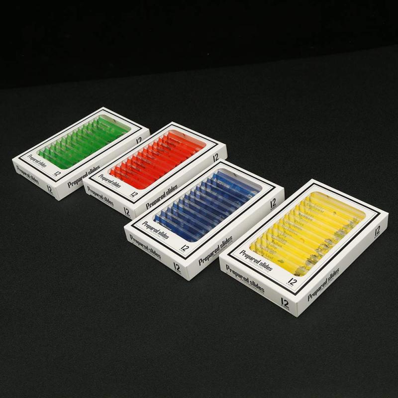
2、 Choosing a stain
How to stain microscope slides:
Staining microscope slides is an essential technique in microscopy that enhances the contrast and visibility of cells and tissues. Here are the steps to follow when staining microscope slides:
1. Prepare the slides: Clean the slides with alcohol and let them dry. Place the specimen on the slide and let it air dry.
2. Choose a stain: There are various types of stains available for different purposes. Some of the commonly used stains include hematoxylin and eosin (H&E), Giemsa, and Wright's stain. The choice of stain depends on the type of specimen and the information required.
3. Apply the stain: Follow the manufacturer's instructions to prepare the stain solution. Apply the stain to the specimen and let it sit for the recommended time. Rinse the slide with distilled water to remove excess stain.
4. Mount the slide: Apply a mounting medium to the slide and cover it with a coverslip. Seal the edges with nail polish or a commercial sealant.
Choosing a stain:
When choosing a stain, it is important to consider the type of specimen and the information required. For example, H&E stain is commonly used to visualize the structure of tissues, while Giemsa stain is used to identify blood cells and parasites. Immunohistochemistry staining is used to detect specific proteins in tissues.
In recent years, there has been a growing interest in using fluorescent dyes for staining microscope slides. Fluorescent dyes offer several advantages over traditional stains, including higher sensitivity and specificity. They are also useful for live cell imaging and can be used to label specific molecules in cells.
In conclusion, staining microscope slides is an essential technique in microscopy that requires careful consideration of the type of stain to use. With the latest advances in staining techniques, researchers can obtain more detailed information about cells and tissues than ever before.

3、 Preparing the stain solution
How to stain microscope slides:
Staining microscope slides is an essential technique in microscopy that enhances the contrast and visibility of cells and tissues. Here are the steps to follow when staining microscope slides:
1. Prepare the slides: Clean the slides thoroughly with soap and water, rinse with distilled water, and dry them with a lint-free cloth.
2. Prepare the specimen: Collect the specimen and prepare it for staining. This may involve fixing the specimen with a fixative solution, such as formalin, to preserve its structure.
3. Preparing the stain solution: There are various types of stains that can be used to stain microscope slides, including hematoxylin and eosin (H&E), Giemsa, and Gram stain. Each stain has a specific protocol for preparation. For example, to prepare a H&E stain, mix hematoxylin and eosin solutions in the appropriate ratio and filter the solution before use.
4. Staining the slides: Place the prepared specimen on the slide and add the stain solution. The staining time varies depending on the type of stain used. After staining, rinse the slide with distilled water and dry it with a lint-free cloth.
5. Mounting the slides: Apply a mounting medium, such as a coverslip, to the slide to protect the specimen and prevent it from drying out.
It is important to note that staining techniques have evolved over time, and new staining methods have been developed to improve the quality of staining and reduce the time required for staining. For example, immunohistochemistry staining uses antibodies to target specific proteins in the specimen, allowing for more precise staining. Additionally, digital staining techniques have been developed that use computer algorithms to simulate the staining process, reducing the need for physical staining.

4、 Applying the stain to the slide
How to stain microscope slides is a crucial technique in the field of microscopy. Staining is used to enhance the contrast of the specimen, making it easier to visualize under the microscope. There are various types of stains available, each with its own specific purpose. Some stains are used to highlight specific structures or organelles, while others are used to differentiate between different types of cells.
To stain a microscope slide, the first step is to prepare the specimen. This involves fixing the specimen onto the slide using a fixative such as formalin or ethanol. Once the specimen is fixed, it can be stained using a variety of techniques.
One common staining technique is the use of dyes such as hematoxylin and eosin (H&E). H&E staining is used to differentiate between different types of cells and tissues. The hematoxylin dye stains the nuclei of cells blue, while the eosin dye stains the cytoplasm and extracellular matrix pink.
Another staining technique is the use of fluorescent dyes. Fluorescent dyes are used to label specific structures or molecules within the specimen. When viewed under a fluorescent microscope, the labeled structures will emit light of a specific wavelength, allowing them to be visualized.
Applying the stain to the slide can be done using a variety of methods. One common method is the use of a staining rack, which allows multiple slides to be stained simultaneously. The staining rack is filled with the staining solution, and the slides are placed into the rack. The staining solution is then allowed to soak into the specimen for a specific amount of time, depending on the type of stain being used.
In conclusion, staining microscope slides is an essential technique in the field of microscopy. There are various types of stains available, each with its own specific purpose. Applying the stain to the slide can be done using a variety of methods, depending on the type of stain being used. With proper staining techniques, it is possible to enhance the contrast of the specimen, making it easier to visualize under the microscope.


