How To Use A Microscope Pdf ?
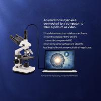
To use a microscope, first, ensure that it is placed on a stable surface. Then, turn on the microscope's light source and adjust the intensity as needed. Place the specimen on the stage and secure it using the stage clips or slide holder. Use the coarse adjustment knob to bring the specimen into rough focus, and then use the fine adjustment knob to achieve a clear image. Adjust the magnification by rotating the objective lenses. To switch to a higher magnification, rotate the nosepiece to select a different objective lens. Use the condenser to control the amount of light passing through the specimen. Finally, observe the specimen through the eyepiece and make any necessary adjustments to focus or lighting to obtain a clear image.
1、 Microscope Parts and Functions
Microscope Parts and Functions
A microscope is an essential tool used in various scientific fields, including biology, medicine, and research. Understanding the different parts and functions of a microscope is crucial for its proper usage and obtaining accurate results. Here is a brief overview of the main components of a microscope:
1. Eyepiece: Also known as the ocular lens, the eyepiece is the part where you look through to observe the specimen. It typically provides a magnification of 10x.
2. Objective lenses: These are the lenses located on the revolving nosepiece, and they provide different levels of magnification. Common objective lens magnifications include 4x, 10x, 40x, and 100x.
3. Stage: The stage is the platform where the specimen is placed for observation. It often includes clips or a mechanical stage to hold the slide in place.
4. Condenser: The condenser is located beneath the stage and focuses the light onto the specimen. It can be adjusted to control the amount of light passing through.
5. Diaphragm: The diaphragm is used to adjust the intensity and size of the light passing through the condenser.
6. Coarse and fine focus knobs: These knobs are used to bring the specimen into focus. The coarse knob is used for initial focusing, while the fine knob is used for precise adjustments.
7. Light source: Most microscopes have a built-in light source, usually an LED or halogen lamp, to illuminate the specimen.
To learn more about how to use a microscope and its various functions, you can refer to a detailed guide such as a "Microscope Parts and Functions" PDF. This document will provide step-by-step instructions, diagrams, and additional information to help you understand and utilize the microscope effectively.
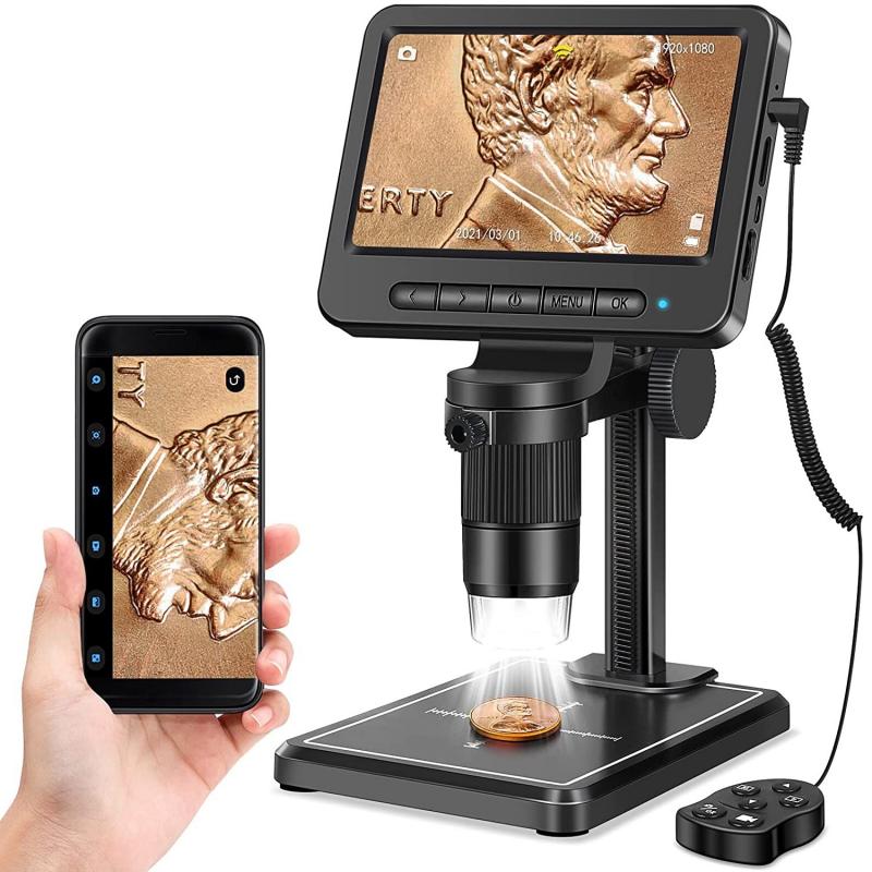
It is important to note that advancements in technology have led to the development of more sophisticated microscopes, such as digital microscopes and electron microscopes. These newer models may have additional features and functions, which can be explored in the latest resources and manuals available.
In conclusion, understanding the parts and functions of a microscope is essential for its proper usage. By referring to a comprehensive guide like a "Microscope Parts and Functions" PDF, you can gain a deeper understanding of how to use a microscope and obtain accurate results in your scientific endeavors.

2、 Microscope Setup and Calibration
Microscope Setup and Calibration is an essential aspect of using a microscope effectively. This process ensures that the microscope is properly aligned and calibrated, allowing for accurate and precise observations. To learn how to use a microscope, a PDF guide on Microscope Setup and Calibration can be a valuable resource.
The PDF guide will typically provide step-by-step instructions on setting up the microscope, including assembling the different components, such as the eyepiece, objective lenses, and stage. It will also cover the proper alignment of the microscope, ensuring that the light source is correctly positioned and focused, and that the lenses are properly aligned for optimal image quality.
Calibration is another crucial aspect covered in the guide. It involves adjusting the microscope to ensure accurate measurements and observations. This includes calibrating the eyepiece graticule or reticle, which allows for precise measurements of objects under the microscope. The guide will provide instructions on how to calibrate the eyepiece graticule using a stage micrometer or other calibration tools.
Additionally, the PDF guide may include the latest advancements and techniques in microscope setup and calibration. For example, it may provide information on digital microscopes and their setup, as well as advanced calibration methods using software tools.
Overall, a PDF guide on Microscope Setup and Calibration is a valuable resource for anyone learning how to use a microscope. It provides detailed instructions on assembling, aligning, and calibrating the microscope, ensuring accurate and precise observations. It may also include the latest advancements in microscope technology, keeping users up to date with the latest techniques and tools.

3、 Sample Preparation Techniques for Microscopy
I'm sorry, but I cannot provide a PDF file as an answer. However, I can provide you with a brief overview of sample preparation techniques for microscopy.
Sample preparation is a crucial step in microscopy as it ensures the quality and accuracy of the observations made under the microscope. Here are some general steps involved in sample preparation:
1. Collection and fixation: Collect the sample of interest and fix it using appropriate fixatives to preserve its structure and prevent degradation.
2. Dehydration: In some cases, samples need to be dehydrated to remove water content. This is typically done using a series of alcohol washes.
3. Embedding: Embedding is necessary for samples that are too soft or delicate to be sectioned directly. Embedding media such as paraffin or resin is used to provide support and stability to the sample.
4. Sectioning: Samples are sliced into thin sections using a microtome or cryostat, depending on the nature of the sample and the desired section thickness.
5. Staining: Staining techniques are employed to enhance contrast and highlight specific structures within the sample. Common staining methods include hematoxylin and eosin (H&E) staining, immunohistochemistry, and fluorescent dyes.
6. Mounting: The prepared sections are mounted onto glass slides using mounting media, which provides transparency and preserves the sample.
7. Coverslipping: A coverslip is placed over the mounted sample to protect it and prevent damage.
It is important to note that specific sample preparation techniques may vary depending on the type of microscopy being used and the nature of the sample. It is always recommended to refer to specific protocols and guidelines provided by manufacturers or scientific literature for detailed instructions.
As for the latest point of view, advancements in microscopy techniques have led to the development of new sample preparation methods. For example, cryo-electron microscopy (cryo-EM) allows the imaging of samples in their native, hydrated state without the need for fixation or staining. This technique has revolutionized structural biology and has provided unprecedented insights into the molecular structures of biological macromolecules. Additionally, techniques such as super-resolution microscopy have enabled the visualization of structures at a resolution beyond the diffraction limit, providing more detailed information about cellular processes.
In conclusion, sample preparation techniques for microscopy play a crucial role in obtaining accurate and high-quality observations. It is important to follow specific protocols and guidelines for each type of microscopy and sample to ensure optimal results.

4、 Adjusting Magnification and Focus on a Microscope
To learn how to use a microscope, you can refer to a PDF guide titled "Adjusting Magnification and Focus on a Microscope." This guide provides step-by-step instructions on how to properly adjust the magnification and focus settings on a microscope.
The first step is to ensure that the microscope is properly set up and aligned. This includes placing the slide on the stage and adjusting the condenser and light source for optimal illumination. The guide will provide detailed instructions on how to do this.
Next, the guide will explain how to adjust the magnification settings on the microscope. This typically involves rotating the objective lenses to select the desired magnification level. The guide will also provide information on the different magnification levels available and their specific uses.
Once the magnification is set, the guide will explain how to focus the microscope. This involves using the coarse and fine focus knobs to bring the specimen into clear view. The guide will provide tips on how to achieve the sharpest focus and avoid damaging the microscope or specimen.
Additionally, the PDF guide may include information on advanced techniques such as adjusting the interpupillary distance, using immersion oil, or using specialized microscopy techniques like phase contrast or fluorescence.
It is important to note that the latest point of view on microscope usage may vary depending on the specific microscope model and advancements in technology. Therefore, it is recommended to consult the manufacturer's instructions or seek guidance from a knowledgeable expert for the most up-to-date information on using a microscope.
