How To Use The Microscope ?
To use a microscope, first, ensure that it is properly set up and adjusted. Place the specimen on the stage and secure it with stage clips. Adjust the focus by using the coarse focus knob to bring the specimen into view, then fine-tune the focus with the fine focus knob. Adjust the magnification by rotating the objective lenses. Use the eyepiece to view the specimen and adjust the focus and magnification as needed. When finished, turn off the microscope and clean the lenses with lens paper.
1、 Microscope Parts and Functions
How to Use the Microscope:
1. Set up the microscope on a stable surface and plug it in if necessary.
2. Adjust the eyepiece to your eye level and focus the objective lens on the lowest magnification setting.
3. Place the specimen on the stage and secure it with the stage clips.
4. Use the coarse adjustment knob to bring the specimen into focus.
5. Adjust the fine adjustment knob to sharpen the image.
6. Increase the magnification by rotating the objective lens to the next level.
7. Use the stage controls to move the specimen around and view different areas.
8. Adjust the diaphragm to control the amount of light entering the microscope.
9. Use the condenser to focus the light on the specimen.
10. Use the eyepiece pointer to mark specific areas of the specimen.
Microscope Parts and Functions:
1. Eyepiece: The lens at the top of the microscope that you look through to view the specimen.
2. Objective Lens: The lens closest to the specimen that magnifies the image.
3. Coarse Adjustment Knob: The knob used to bring the specimen into focus.
4. Fine Adjustment Knob: The knob used to sharpen the image.
5. Stage: The platform where the specimen is placed.
6. Stage Clips: The clips that hold the specimen in place on the stage.
7. Diaphragm: The part that controls the amount of light entering the microscope.
8. Condenser: The lens that focuses the light on the specimen.
9. Light Source: The part that provides the light for viewing the specimen.
10. Base: The bottom of the microscope that provides stability.
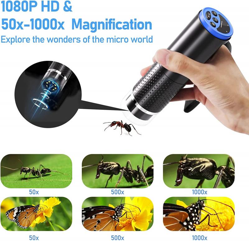
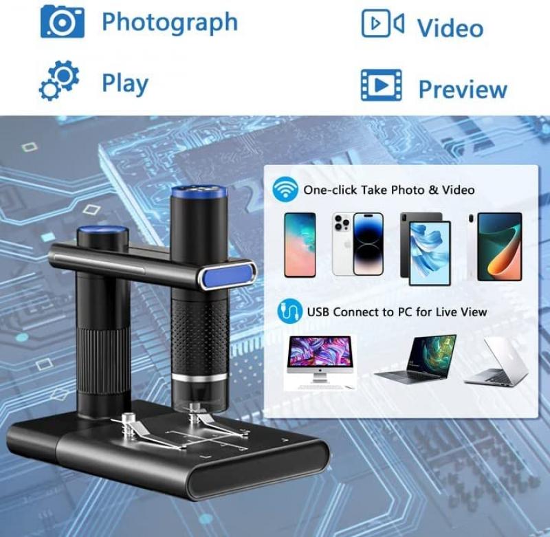
In addition to these traditional parts and functions, modern microscopes may also include digital imaging capabilities, allowing for the capture and analysis of images on a computer. Some microscopes may also have advanced features such as fluorescence imaging, which allows for the visualization of specific molecules within a specimen. As technology continues to advance, the capabilities of microscopes will continue to expand, allowing for even more detailed and precise observations of the microscopic world.
2、 Microscope Preparation Techniques
How to use the microscope:
1. Familiarize yourself with the microscope: Before using the microscope, it is important to familiarize yourself with its parts and functions. This includes the eyepiece, objective lenses, stage, focus knobs, and light source.
2. Prepare your sample: Microscope preparation techniques vary depending on the type of sample being observed. For example, samples may need to be stained or mounted on a slide. It is important to follow proper preparation techniques to ensure accurate observations.
3. Place the sample on the stage: Once your sample is prepared, place it on the stage of the microscope. Use the stage clips to secure the sample in place.
4. Adjust the focus: Use the coarse focus knob to bring the sample into focus. Once the sample is roughly in focus, use the fine focus knob to make small adjustments and achieve a clear image.
5. Select the appropriate objective lens: Microscopes typically have multiple objective lenses with varying magnification levels. Select the appropriate lens for your sample and adjust the focus as needed.
6. Observe and record your findings: Once your sample is in focus, observe and record your findings. This may include taking measurements or capturing images using a camera attached to the microscope.
It is important to note that the use of microscopes has evolved over time with the advancement of technology. For example, digital microscopes now allow for real-time observation and recording of samples, while confocal microscopy allows for 3D imaging of samples. It is important to stay up-to-date with the latest techniques and technologies in order to make the most accurate observations and interpretations.
3、 Microscope Illumination Methods
Microscope Illumination Methods:
Microscope illumination is an essential aspect of microscopy, as it determines the quality of the image produced. There are several illumination methods used in microscopy, including brightfield, darkfield, phase contrast, and fluorescence microscopy.
1. Brightfield microscopy: This is the most common illumination method used in microscopy. It involves illuminating the sample with a bright light source from below the stage. The light passes through the sample and is then magnified by the objective lens. This method is ideal for observing stained samples, as the contrast between the sample and the background is enhanced.
2. Darkfield microscopy: This method involves illuminating the sample with a hollow cone of light, which is directed at an angle to the sample. The light is scattered by the sample, producing a bright image against a dark background. This method is ideal for observing unstained samples, such as live bacteria, as it enhances the contrast between the sample and the background.
3. Phase contrast microscopy: This method involves using a special condenser and objective lens to produce contrast in unstained samples. The condenser produces a phase shift in the light passing through the sample, which is then detected by the objective lens. This method is ideal for observing live cells, as it produces high contrast images without the need for staining.
4. Fluorescence microscopy: This method involves using a fluorescent dye to label specific structures within the sample. The sample is then illuminated with a specific wavelength of light, which causes the dye to emit light of a different color. This method is ideal for observing specific structures within the sample, such as DNA or proteins.
In conclusion, the choice of illumination method depends on the type of sample being observed and the information required. The latest point of view is that advances in technology have led to the development of new illumination methods, such as super-resolution microscopy, which allows for the observation of structures at the nanoscale level.

4、 Microscope Magnification and Resolution
How to use the microscope:
1. Familiarize yourself with the parts of the microscope: The microscope has several parts, including the eyepiece, objective lenses, stage, and focus knobs. Understanding the function of each part is essential to using the microscope effectively.
2. Prepare your sample: Place your sample on the stage and secure it in place using the stage clips. Adjust the position of the sample so that it is in the center of the field of view.
3. Adjust the focus: Use the coarse focus knob to bring the sample into focus. Once the sample is roughly in focus, use the fine focus knob to adjust the focus more precisely.
4. Choose the appropriate objective lens: The microscope has several objective lenses with different magnifications. Choose the appropriate lens for your sample and adjust the magnification as needed.
5. Observe and record your findings: Once your sample is in focus, observe it carefully and record your observations. You may need to adjust the focus or magnification as you observe different parts of the sample.
Microscope Magnification and Resolution:
Microscope magnification refers to the degree to which the image of a sample is enlarged. The higher the magnification, the more detail you can see. However, higher magnification also means a smaller field of view and a shallower depth of field.
Resolution refers to the ability of the microscope to distinguish between two closely spaced objects. The higher the resolution, the more detail you can see in the image. However, resolution is limited by the wavelength of light used to illuminate the sample.
Recent advances in microscopy technology have led to the development of super-resolution microscopy techniques, which allow for even higher levels of magnification and resolution. These techniques use specialized equipment and software to overcome the limitations of traditional microscopy and provide unprecedented detail and clarity in imaging.


