How To View Bacteria Under Microscope ?
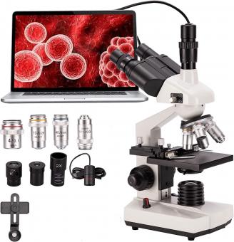
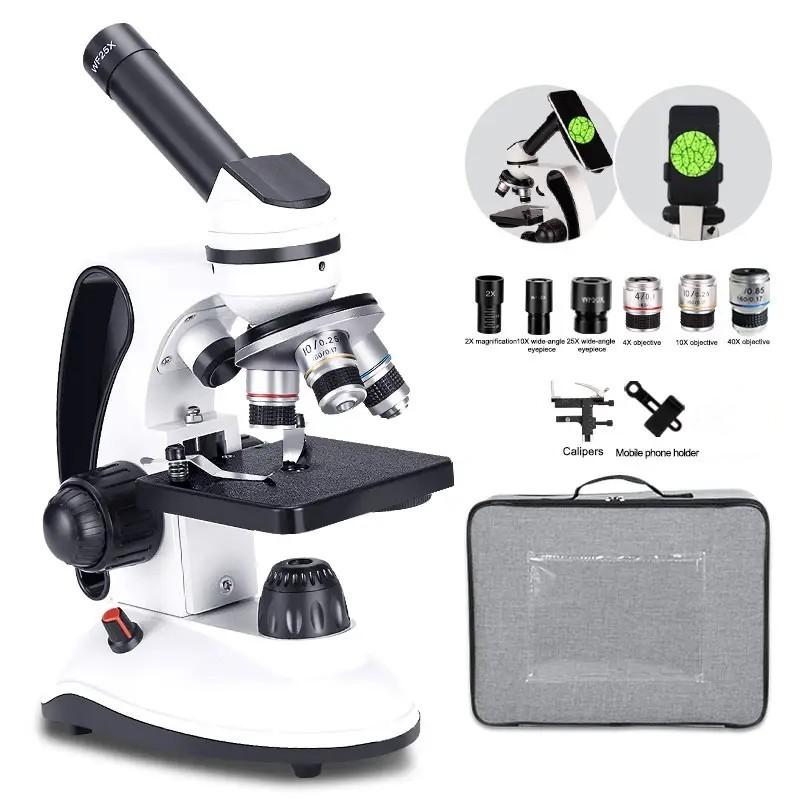
To view bacteria under a microscope, you will need a compound light microscope with a high magnification power. Start by preparing a bacterial sample by taking a small amount of the bacteria culture and placing it on a clean microscope slide. Add a drop of water or a special staining solution to the sample to enhance visibility. Gently place a coverslip over the sample to avoid air bubbles.
Next, adjust the microscope's focus by starting with the lowest magnification objective lens and gradually increasing the magnification until the bacteria come into view. Use the fine focus knob to sharpen the image. It may be helpful to use the condenser to adjust the amount of light passing through the sample.
Once the bacteria are visible, observe their shape, size, and movement. You can also take photographs or make drawings to document your observations. Remember to properly clean and store the microscope after use.
1、 Microscope Preparation Techniques for Bacterial Observation
Microscope Preparation Techniques for Bacterial Observation
To view bacteria under a microscope, proper preparation techniques are essential to ensure accurate observation and analysis. Here is a step-by-step guide on how to prepare bacterial samples for microscopic observation:
1. Sample Collection: Collect a small sample of the bacteria you wish to observe. This can be obtained from various sources such as water, soil, or a culture medium.
2. Fixation: Fixation is crucial to preserve the bacterial structure and prevent distortion during the observation process. Common fixatives include heat, alcohol, or formaldehyde. The choice of fixative depends on the specific requirements of the experiment.
3. Staining: Staining is used to enhance the visibility of bacteria under the microscope. The most commonly used staining techniques are Gram staining and acid-fast staining. These techniques help differentiate bacteria based on their cell wall composition and aid in their identification.
4. Mounting: After staining, the bacteria need to be mounted on a slide for observation. A drop of the stained bacterial suspension is placed on a clean glass slide, covered with a coverslip, and sealed with a mounting medium to prevent drying.
5. Microscopic Observation: Place the prepared slide on the microscope stage and start with the lowest magnification objective lens. Gradually increase the magnification to observe the bacteria in detail. Adjust the focus and lighting as necessary to obtain a clear image.
It is important to note that advancements in microscopy techniques have led to the development of more sophisticated methods for bacterial observation. For instance, fluorescence microscopy allows the visualization of specific bacterial components or proteins by using fluorescent dyes or antibodies. Additionally, confocal microscopy provides three-dimensional imaging of bacterial structures, enabling a more comprehensive understanding of their organization and behavior.
In conclusion, proper preparation techniques, including fixation, staining, and mounting, are crucial for observing bacteria under a microscope. The choice of staining technique and the use of advanced microscopy methods can further enhance the accuracy and depth of bacterial observation.
2、 Staining Methods for Enhanced Bacterial Visualization
To view bacteria under a microscope, staining methods are commonly used to enhance their visualization. Staining techniques involve the application of dyes or stains to bacterial cells, which helps to highlight their structures and make them more visible under the microscope. There are several staining methods that can be employed for this purpose.
One commonly used staining method is the Gram stain, which differentiates bacteria into two groups: Gram-positive and Gram-negative. This technique involves the application of crystal violet, iodine, alcohol, and safranin to the bacterial cells. Gram-positive bacteria retain the crystal violet stain, appearing purple, while Gram-negative bacteria lose the stain and appear pink after the application of safranin.
Another staining method is the acid-fast stain, which is used specifically for the identification of Mycobacterium species, including the causative agent of tuberculosis. Acid-fast staining involves the application of carbol fuchsin, acid-alcohol, and methylene blue to the bacterial cells. Acid-fast bacteria retain the carbol fuchsin stain even after the application of acid-alcohol, while other bacteria lose the stain and appear blue after the application of methylene blue.
Fluorescent staining is another technique that has gained popularity in recent years. This method involves the use of fluorescent dyes that bind specifically to certain bacterial structures or components, such as DNA or cell membranes. The stained bacteria can then be visualized using a fluorescence microscope, which emits light of a specific wavelength to excite the fluorescent dyes and produce a bright image.
It is important to note that the choice of staining method depends on the specific requirements of the study or experiment. Each staining technique has its advantages and limitations, and researchers should carefully select the appropriate method based on the bacteria being studied and the information sought.
In conclusion, staining methods are essential for enhancing the visualization of bacteria under a microscope. Techniques such as Gram staining, acid-fast staining, and fluorescent staining provide valuable insights into the structure and characteristics of bacterial cells. Researchers continue to explore and develop new staining methods to improve bacterial visualization and gain a deeper understanding of these microorganisms.

3、 Adjusting Microscope Settings for Bacterial Observation
To view bacteria under a microscope, it is essential to adjust the microscope settings appropriately. Here is a step-by-step guide on how to do so:
1. Start by preparing a bacterial sample. This can be done by taking a small amount of the bacteria culture and placing it on a clean microscope slide. Ensure that the slide is clean and free from any debris.
2. Once the sample is prepared, place the slide on the microscope stage and secure it using the stage clips. Make sure the sample is centered and in focus.
3. Begin by using the lowest magnification objective lens (usually 4x or 10x) and adjust the coarse focus knob to bring the sample into rough focus. Use the fine focus knob to further refine the focus.
4. Once the sample is in focus, increase the magnification by rotating the nosepiece to a higher objective lens (40x or 100x). Use the fine focus knob again to bring the sample into sharp focus.
5. Adjust the condenser height to ensure proper illumination. The condenser should be close to the slide but not touching it. Open the diaphragm to allow more light to pass through the sample.
6. If necessary, adjust the brightness and contrast settings on the microscope to enhance the visibility of the bacteria. This can be done using the microscope's built-in controls or by adjusting the lighting conditions in the room.
7. Finally, observe the sample through the eyepiece and use the stage controls to move the slide and explore different areas. Take note of the size, shape, and arrangement of the bacteria.
It is important to note that advancements in microscopy techniques, such as fluorescence microscopy and electron microscopy, have allowed for more detailed and specific observations of bacteria. These techniques can provide insights into the internal structures and functions of bacteria, aiding in research and diagnostics.

4、 Identifying Bacterial Morphology and Cellular Structures
To view bacteria under a microscope, follow these steps:
1. Prepare a bacterial sample: Collect a small amount of the bacteria you want to view. This can be obtained from a culture plate or a liquid culture. Ensure that the sample is pure and free from contaminants.
2. Prepare a microscope slide: Place a drop of water or saline solution on a clean microscope slide. Using a sterile loop or pipette, transfer a small amount of the bacterial sample onto the slide. Spread the sample evenly across the slide.
3. Stain the bacteria: Staining helps to enhance the visibility of bacteria under the microscope. The most commonly used stain is the Gram stain, which differentiates bacteria into Gram-positive and Gram-negative based on their cell wall composition. Other stains, such as methylene blue or crystal violet, can also be used.
4. Cover the slide: Place a coverslip over the stained bacterial sample. Gently press down to remove any air bubbles.
5. Set up the microscope: Place the slide on the microscope stage and secure it with the stage clips. Start with the lowest magnification objective lens and gradually increase the magnification until the bacteria are visible.
6. Focus and observe: Use the coarse and fine adjustment knobs to bring the bacteria into focus. Observe the bacterial morphology, such as shape (rod, cocci, spiral), arrangement (clusters, chains, pairs), and size. Additionally, observe any cellular structures, such as flagella or capsules, if present.
It is important to note that advancements in microscopy techniques, such as fluorescence microscopy and electron microscopy, have allowed for more detailed visualization of bacterial structures. These techniques can provide insights into the internal organization of bacteria and the localization of specific proteins or molecules within the cells. However, these advanced techniques require specialized equipment and expertise.
