Light Microscope How It Works ?
A light microscope works by using visible light to illuminate a specimen and magnify it for observation. The light source, usually a bulb, emits light that passes through a condenser lens, which focuses the light onto the specimen. The light then passes through the objective lens, which further magnifies the image. The objective lens is located close to the specimen and collects the light that is transmitted through it. The magnified image is then projected through the eyepiece lens, which further enlarges the image for the viewer to see. The eyepiece lens is located at the top of the microscope and is where the viewer looks into the microscope. By adjusting the focus knobs, the viewer can bring the specimen into sharp focus. Light microscopes are commonly used in various scientific fields, such as biology and medicine, to study small organisms, cells, and tissues.
1、 Optical principles of light microscopy
The light microscope, also known as an optical microscope, is a widely used tool in scientific research and medical diagnostics. It works on the principles of optics to magnify and visualize small objects that are otherwise invisible to the naked eye.
The basic working principle of a light microscope involves the use of visible light to illuminate the specimen and a series of lenses to magnify the image. The light source, typically a halogen lamp, emits light that passes through a condenser lens, which focuses the light onto the specimen. The light then passes through the objective lens, which further magnifies the image, and finally through the eyepiece lens, which allows the viewer to observe the magnified image.
The magnification power of a light microscope is determined by the combination of the objective lens and the eyepiece lens. The objective lens is responsible for the primary magnification, typically ranging from 4x to 100x, while the eyepiece lens further magnifies the image, usually by 10x. This results in a total magnification of up to 1000x.
In addition to magnification, light microscopes also utilize various techniques to enhance contrast and improve image quality. These techniques include phase contrast, differential interference contrast, and fluorescence microscopy, among others. These methods allow for the visualization of specific structures or molecules within the specimen.
The latest advancements in light microscopy include the development of super-resolution techniques, such as stimulated emission depletion (STED) microscopy and structured illumination microscopy (SIM). These techniques surpass the traditional diffraction limit of light, enabling the visualization of structures at the nanoscale level.
Overall, the light microscope continues to be a fundamental tool in scientific research, providing valuable insights into the microscopic world and contributing to advancements in various fields, including biology, medicine, and materials science.

2、 Components and functions of a light microscope
A light microscope is an essential tool used in various scientific fields to observe and study microscopic organisms and structures. It works by using visible light to magnify and illuminate the specimen being observed.
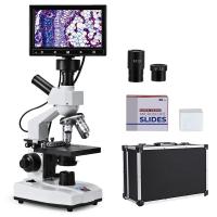
The main components of a light microscope include the eyepiece, objective lenses, stage, condenser, and light source. The eyepiece is where the observer looks through to view the specimen. The objective lenses, usually ranging from 4x to 100x magnification, are responsible for magnifying the image. The stage holds the specimen in place, and the condenser focuses the light onto the specimen. The light source, typically a bulb, provides the necessary illumination.
When using a light microscope, the specimen is placed on the stage and adjusted to the desired magnification using the objective lenses. The light source is then turned on, and the condenser focuses the light onto the specimen. The light passes through the specimen, and some of it is absorbed while the rest is transmitted. The transmitted light is then collected by the objective lenses and further magnified. Finally, the image is viewed through the eyepiece, allowing the observer to see the magnified specimen.
Advancements in light microscopy have led to the development of techniques such as fluorescence microscopy, confocal microscopy, and super-resolution microscopy. These techniques utilize fluorescent dyes, laser scanning, and complex imaging algorithms to enhance the resolution and contrast of the images obtained. Additionally, digital imaging technology has allowed for the capture and analysis of microscope images using computer software, enabling further analysis and documentation of the observed specimens.
In conclusion, a light microscope works by using visible light to magnify and illuminate a specimen. Its components, such as the eyepiece, objective lenses, stage, condenser, and light source, work together to produce a magnified image that can be observed and studied. Ongoing advancements in light microscopy continue to improve the resolution and capabilities of this essential scientific tool.

3、 Illumination techniques in light microscopy
The light microscope is a widely used tool in biological research and allows scientists to observe and study cells and tissues at a microscopic level. Understanding how it works is crucial for obtaining clear and accurate images.
The basic principle of a light microscope involves the use of visible light to illuminate the specimen and magnify the image. The light source, typically a halogen lamp, emits light that passes through a condenser lens. The condenser focuses the light onto the specimen, which is placed on a glass slide. The light then passes through the objective lens, which further magnifies the image, and finally through the eyepiece, where it is observed by the researcher.
Illumination techniques play a crucial role in enhancing the contrast and resolution of the specimen. One commonly used technique is brightfield microscopy, where the specimen is observed against a bright background. This technique is suitable for observing stained samples, but it may lack contrast for unstained specimens.
To overcome this limitation, various contrast-enhancing techniques have been developed. Phase contrast microscopy, for example, exploits the differences in refractive index between the specimen and its surrounding medium to create contrast. Differential interference contrast (DIC) microscopy uses polarized light to create a 3D-like image with enhanced contrast.
Fluorescence microscopy is another powerful technique that utilizes fluorescent dyes or proteins to label specific structures within the specimen. The specimen is illuminated with a specific wavelength of light, and the emitted fluorescence is captured and visualized. This technique allows for the visualization of specific molecules or structures within cells, providing valuable insights into their function and localization.
In recent years, advancements in technology have led to the development of super-resolution microscopy techniques, such as stimulated emission depletion (STED) microscopy and structured illumination microscopy (SIM). These techniques surpass the diffraction limit of light, enabling the visualization of structures at a resolution previously thought impossible.
In conclusion, the light microscope works by illuminating the specimen with visible light and magnifying the image using lenses. Various illumination techniques, such as brightfield, phase contrast, and fluorescence microscopy, enhance the contrast and resolution of the specimen. The latest advancements in super-resolution microscopy have revolutionized the field, allowing for the visualization of structures at an unprecedented level of detail.

4、 Image formation and magnification in light microscopy
A light microscope works by using visible light to illuminate a sample and produce an image. The basic principle of image formation in light microscopy involves the interaction of light with the sample and the subsequent magnification of the image.
When light passes through the sample, it undergoes various interactions such as absorption, reflection, and scattering. These interactions depend on the properties of the sample, such as its composition and structure. The light that is transmitted through or reflected by the sample then enters the objective lens of the microscope.
The objective lens collects the light and focuses it onto the intermediate image plane, also known as the image plane. This is where the image of the sample is formed. The objective lens plays a crucial role in determining the resolution and magnification of the microscope. It has a high numerical aperture, which allows it to collect light from a wide range of angles and improve the resolution of the image.
The image formed at the image plane is then magnified by the eyepiece lens, which further enlarges the image and allows the viewer to observe it. The total magnification of the microscope is the product of the magnification of the objective lens and the eyepiece lens.
In recent years, advancements in light microscopy have led to the development of techniques such as confocal microscopy and super-resolution microscopy. Confocal microscopy uses a pinhole to eliminate out-of-focus light, resulting in improved image quality and resolution. Super-resolution microscopy techniques, such as stimulated emission depletion (STED) microscopy and structured illumination microscopy (SIM), surpass the diffraction limit of light and enable the visualization of structures at the nanoscale.
Overall, light microscopy continues to be a powerful tool in biological research, allowing scientists to observe and study a wide range of samples with high resolution and magnification.


There are no comments for this blog.