What Are All The Parts Of A Microscope?
A microscope typically consists of several parts, including the eyepiece, objective lenses, stage, focus knobs, illuminator, and base. The eyepiece is the lens that the viewer looks through to see the magnified image. The objective lenses are the lenses closest to the specimen and are responsible for magnifying the image. The stage is the platform on which the specimen is placed for observation. The focus knobs are used to adjust the focus of the image. The illuminator provides light to illuminate the specimen. The base is the bottom part of the microscope that provides stability and support.
1、 Eyepiece

The microscope is an essential tool in the field of science, allowing us to observe and study objects that are too small to be seen with the naked eye. It is composed of several parts that work together to magnify and illuminate the specimen being observed.
The eyepiece, also known as the ocular lens, is the part of the microscope that the viewer looks through to observe the specimen. It typically has a magnification power of 10x and is located at the top of the microscope.
Other important parts of the microscope include the objective lenses, which are located on the revolving nosepiece and provide varying levels of magnification. The stage is where the specimen is placed for observation, and it often includes clips or other mechanisms to hold the specimen in place.
The condenser is a lens system located beneath the stage that focuses the light onto the specimen, while the diaphragm controls the amount of light that passes through the condenser. The light source, typically an LED or halogen bulb, provides illumination for the specimen.
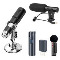
In recent years, digital microscopes have become increasingly popular, allowing for the capture and analysis of images using a camera and computer software. These microscopes often include additional parts such as a USB camera, software for image analysis, and a monitor for viewing the specimen.
Overall, the microscope is a complex instrument with many parts that work together to provide a detailed view of the microscopic world. As technology continues to advance, we can expect to see further developments in the design and functionality of microscopes.
2、 Objective lens
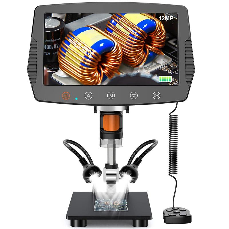
What are all the parts of a microscope? A microscope is an essential tool used in scientific research, medical diagnosis, and education. It is a device that magnifies small objects, making them visible to the human eye. The microscope has several parts that work together to produce a clear and magnified image of the specimen being observed.
The main parts of a microscope include the eyepiece, objective lens, stage, focus knobs, and illuminator. The eyepiece is the part of the microscope that the viewer looks through to see the magnified image. The objective lens is the lens closest to the specimen and is responsible for magnifying the image. The stage is the platform on which the specimen is placed for observation. The focus knobs are used to adjust the focus of the image, and the illuminator provides light to the specimen.
In recent years, there have been advancements in microscope technology, leading to the development of new parts and features. For example, some microscopes now come with digital cameras that allow for the capture and storage of images. Additionally, some microscopes have computer software that can analyze and measure the images captured.
In conclusion, the parts of a microscope are essential for producing clear and magnified images of small objects. As technology advances, new parts and features are being added to microscopes, making them even more useful in scientific research, medical diagnosis, and education.
3、 Stage

What are all the parts of a microscope? A microscope is an essential tool used in scientific research, medical diagnosis, and education. It is a device that magnifies small objects, making them visible to the human eye. The microscope has several parts that work together to produce a clear and magnified image of the specimen being observed.
The main parts of a microscope include the eyepiece, objective lenses, stage, focus knobs, and light source. The eyepiece is the part of the microscope that the viewer looks through to see the magnified image. The objective lenses are located on the nosepiece and are used to magnify the specimen. The stage is the platform on which the specimen is placed for observation. The focus knobs are used to adjust the focus of the microscope, making the image clearer and sharper. The light source illuminates the specimen, making it easier to see.
In recent years, there have been advancements in microscope technology, leading to the development of new parts and features. For example, some microscopes now come with digital cameras that allow users to capture images and videos of the specimen being observed. Additionally, some microscopes have built-in software that can analyze and measure the specimen automatically.
In conclusion, the parts of a microscope are essential for producing a clear and magnified image of the specimen being observed. With advancements in technology, new parts and features have been added to microscopes, making them even more useful in scientific research, medical diagnosis, and education.
4、 Diaphragm
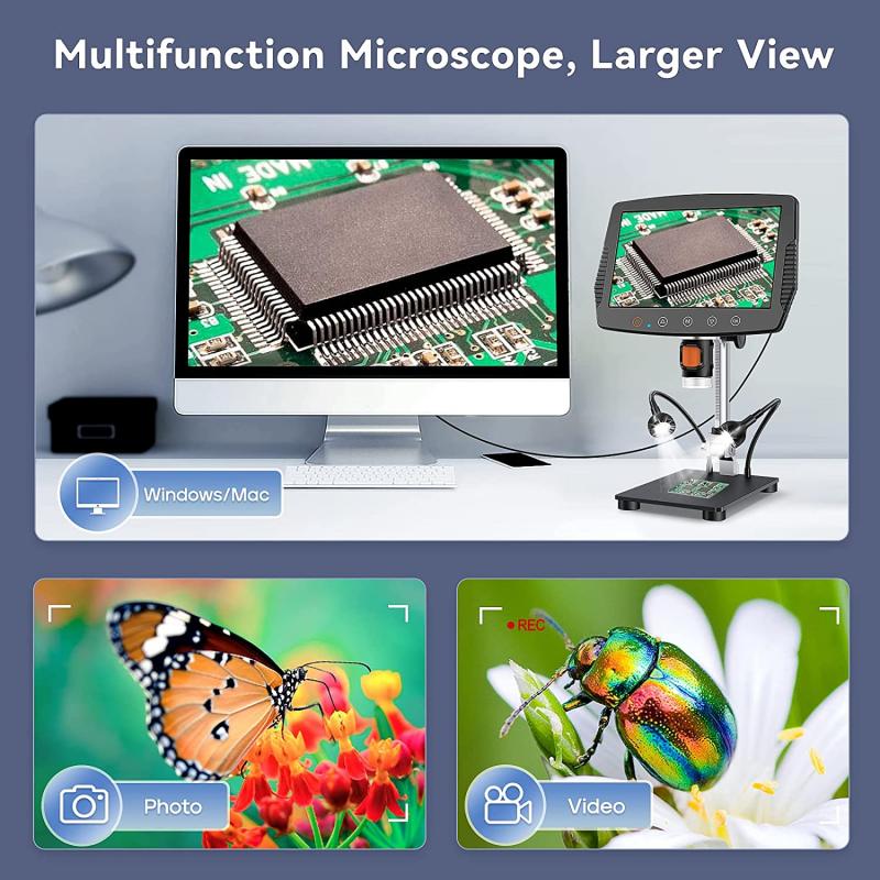
The microscope is an essential tool in the field of science, allowing us to observe and study objects that are too small to be seen with the naked eye. A microscope consists of several parts that work together to magnify and illuminate the specimen being observed.
The main parts of a microscope include the eyepiece, objective lenses, stage, focus knobs, and light source. The eyepiece is the lens that the viewer looks through to observe the specimen. The objective lenses are the lenses closest to the specimen and are responsible for magnifying the image. The stage is the platform on which the specimen is placed for observation. The focus knobs are used to adjust the focus of the image, and the light source illuminates the specimen.
Another important part of a microscope is the diaphragm, which controls the amount of light that passes through the specimen. By adjusting the diaphragm, the viewer can control the brightness and contrast of the image, making it easier to observe and study.
In recent years, advances in technology have led to the development of digital microscopes, which use cameras and computer software to capture and analyze images. These microscopes have additional parts, such as a digital camera and software for image processing and analysis.
Overall, the parts of a microscope work together to provide a clear and detailed view of the microscopic world, allowing scientists to make important discoveries and advancements in various fields of study.


