What Are The Different Magnifications On A Microscope ?
Microscopes have different magnifications that allow users to view objects at different levels of detail. The magnification of a microscope is determined by the combination of lenses in the eyepiece and objective lenses. The most common magnifications on a microscope are:
1. Low power (10x-40x): This is the lowest magnification on a microscope and is used to view larger specimens such as whole organisms or tissues.
2. Medium power (100x-400x): This magnification is used to view smaller specimens such as cells and bacteria.
3. High power (400x-1000x): This magnification is used to view very small specimens such as individual cells and cell structures.
4. Oil immersion (1000x-2000x): This is the highest magnification on a microscope and is used to view very small structures such as viruses and subcellular structures. It requires the use of a special oil to improve the resolution of the image.
1、 Low power magnification

The different magnifications on a microscope typically include low power, medium power, and high power magnification. Low power magnification is usually the lowest magnification setting on a microscope and is used to view larger specimens or to get an overall view of the specimen. This magnification is typically between 10x and 40x, depending on the microscope.
Medium power magnification is the next level of magnification and is used to view smaller details of the specimen. This magnification is typically between 100x and 400x, depending on the microscope. High power magnification is the highest magnification setting on a microscope and is used to view the smallest details of the specimen. This magnification is typically between 400x and 1000x, depending on the microscope.
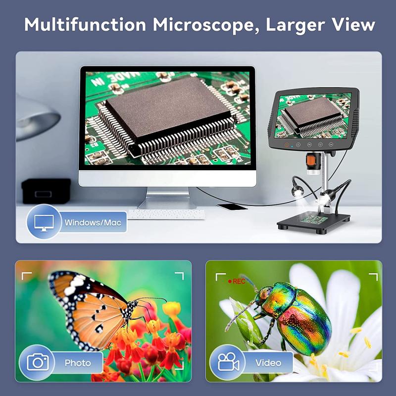
In recent years, there has been a trend towards using digital microscopes that allow for even higher magnifications and more detailed imaging. These microscopes use digital cameras and software to capture and analyze images, allowing for greater precision and accuracy in scientific research and medical diagnosis.
Overall, the different magnifications on a microscope are essential for viewing and analyzing specimens at different levels of detail. From low power magnification to high power magnification, each level provides a unique perspective on the specimen and helps scientists and researchers gain a better understanding of the world around us.
2、 Medium power magnification

The different magnifications on a microscope typically include low power, medium power, and high power magnification. Low power magnification is usually around 40x to 100x, medium power magnification is around 100x to 400x, and high power magnification is around 400x to 1000x or more. However, the exact magnification range can vary depending on the type of microscope and the lenses used.
Medium power magnification is particularly useful for observing cellular structures and small organisms. At this magnification, it is possible to see individual cells and their organelles, as well as bacteria and other microorganisms. This level of magnification is often used in medical research and diagnosis, as well as in biology and microbiology studies.
In recent years, advances in technology have led to the development of new types of microscopes with even higher magnification capabilities. For example, electron microscopes can magnify objects up to 1,000,000x, allowing for the observation of even smaller structures such as viruses and molecules. Additionally, confocal microscopes use lasers to create 3D images of cells and tissues, providing a more detailed view of their internal structures.
Overall, the different magnifications on a microscope allow scientists and researchers to observe and study the world at a microscopic level, providing valuable insights into the workings of living organisms and the natural world.
3、 High power magnification
The different magnifications on a microscope typically include low power, medium power, and high power magnification. Low power magnification is usually around 40x to 100x, and is used to view larger specimens such as whole organisms or tissues. Medium power magnification is typically around 100x to 400x, and is used to view smaller structures such as cells and cell organelles. High power magnification is usually around 400x to 1000x, and is used to view even smaller structures such as bacteria and viruses.
However, with the advancement of technology, microscopes with higher magnification capabilities have been developed. For example, electron microscopes can magnify specimens up to 2 million times, allowing for the visualization of even smaller structures such as molecules and atoms. These microscopes use beams of electrons instead of light to create an image, and require specialized techniques for sample preparation.
In addition to magnification, microscopes can also have different types of lenses and illumination systems, which can affect the quality and clarity of the image. Some microscopes also have additional features such as digital imaging capabilities, which allow for the capture and analysis of images using computer software.
Overall, the different magnifications on a microscope are important for studying and understanding the structures and functions of various biological specimens. As technology continues to advance, we can expect even higher magnification capabilities and more advanced imaging techniques to further enhance our understanding of the microscopic world.
4、 Oil immersion magnification

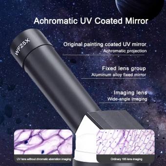
The different magnifications on a microscope typically range from 40x to 1000x, depending on the type of microscope and the lenses used. The magnification power of a microscope is determined by the combination of the objective lens and the eyepiece lens. The objective lens is the lens closest to the specimen, while the eyepiece lens is the lens closest to the eye.
The magnification power of a microscope can be increased by using oil immersion. Oil immersion is a technique used to increase the resolution and magnification of a microscope. It involves placing a drop of oil on the specimen and using a special lens with a high numerical aperture to view the specimen. The oil has the same refractive index as the lens, which reduces the amount of light that is lost and increases the resolution of the image.
Oil immersion magnification can be used in a variety of applications, including medical diagnosis, research, and education. It is particularly useful in the study of bacteria and other microorganisms, as it allows for a more detailed examination of their structure and behavior.
In recent years, advances in technology have led to the development of new types of microscopes with even higher magnification powers. For example, electron microscopes use beams of electrons instead of light to magnify specimens, allowing for magnifications of up to 10 million times. These high-powered microscopes have revolutionized the field of microscopy and have opened up new avenues of research in fields such as nanotechnology and materials science.


