What Are The Parts Of A Compound Microscope ?
The main parts of a compound microscope include the eyepiece, objective lenses, stage, condenser, diaphragm, focus knobs, and light source.
1、 Eyepiece
The compound microscope is a powerful tool used in scientific research, education, and various other fields. It consists of several essential parts that work together to magnify and visualize microscopic specimens. The main parts of a compound microscope include the eyepiece, objective lenses, stage, condenser, focus knobs, and illuminator.
The eyepiece, also known as the ocular lens, is the part where the viewer looks through to observe the specimen. It typically provides a magnification of 10x, although higher magnification eyepieces are also available. The eyepiece is usually adjustable to accommodate different users' eyesight.
The objective lenses are located on a rotating nosepiece and are responsible for magnifying the specimen. Compound microscopes typically have multiple objective lenses with different magnification powers, such as 4x, 10x, 40x, and 100x. By rotating the nosepiece, different objective lenses can be selected for observation.
The stage is a flat platform where the specimen is placed for examination. It often includes clips or mechanical stage controls to secure and move the specimen smoothly. The stage may also have a stage micrometer, which is a calibrated ruler used for measuring the size of the specimen.
The condenser is located beneath the stage and helps focus the light onto the specimen. It consists of lenses that concentrate and direct the light, enhancing the clarity and brightness of the image. The condenser can be adjusted to control the amount of light reaching the specimen.
The focus knobs are used to bring the specimen into sharp focus. Most compound microscopes have two focus knobs, one for coarse adjustment and another for fine adjustment. The coarse adjustment knob is used to quickly bring the specimen into approximate focus, while the fine adjustment knob is used for precise focusing.
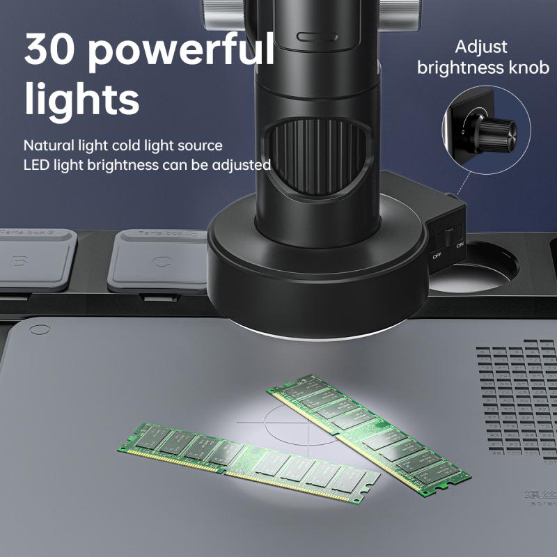
The illuminator is the light source of the compound microscope. It can be built-in or external, and it provides the necessary illumination to visualize the specimen. Modern microscopes often use LED lights as they are energy-efficient and provide consistent lighting.
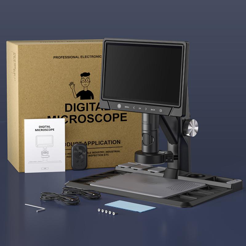
In recent years, advancements in technology have led to the development of digital compound microscopes. These microscopes incorporate a camera and a screen, allowing users to view and capture images or videos of the specimen directly. This digital capability has revolutionized microscopy, enabling easier sharing of observations and analysis.
In conclusion, the compound microscope consists of several essential parts, including the eyepiece, objective lenses, stage, condenser, focus knobs, and illuminator. These components work together to magnify and visualize microscopic specimens, aiding in scientific research, education, and various other applications. With the advent of digital technology, microscopes have become even more versatile and accessible, opening up new possibilities in the field of microscopy.
2、 Objective lenses
The compound microscope is a powerful tool used in scientific research, education, and various other fields. It consists of several essential parts that work together to magnify and visualize microscopic specimens. One of the key components of a compound microscope is the objective lenses.
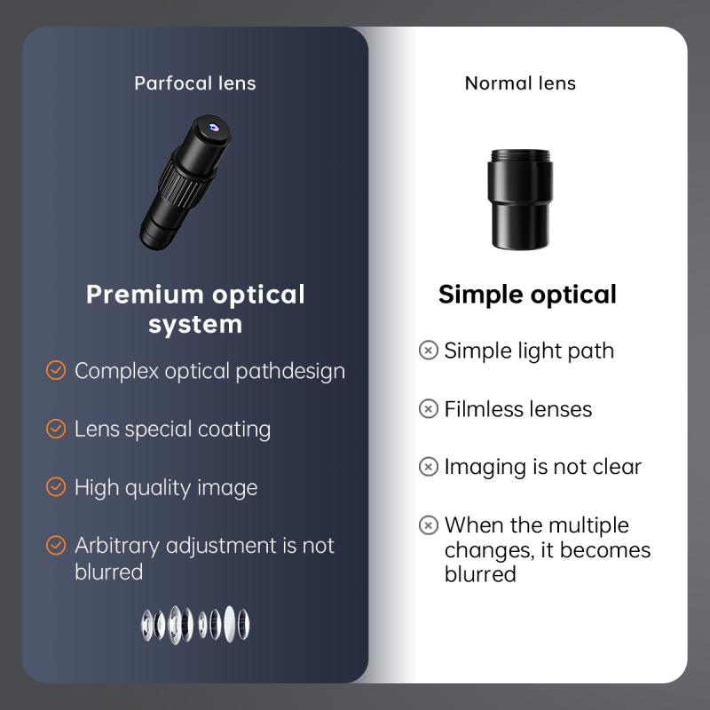
Objective lenses are a set of lenses located at the lower end of the microscope's body tube. They are responsible for gathering light from the specimen and magnifying it to produce a clear and detailed image. Compound microscopes typically have multiple objective lenses with different magnification powers, allowing users to observe specimens at various levels of detail.
The objective lenses are usually color-coded or labeled with numbers to indicate their magnification power. Common magnification powers include 4x (low power), 10x (medium power), 40x (high power), and 100x (oil immersion). The magnification power of the objective lenses, combined with the eyepiece lenses, determines the total magnification of the microscope.
In recent years, advancements in technology have led to the development of specialized objective lenses. For example, there are now objective lenses designed specifically for fluorescence microscopy, which allow researchers to observe fluorescently labeled specimens with high sensitivity and resolution. Additionally, there are objective lenses with correction features to compensate for aberrations and improve image quality.
Objective lenses are a critical component of a compound microscope, enabling scientists and researchers to explore the microscopic world with precision and clarity. As technology continues to advance, we can expect further improvements in objective lens design, leading to even more powerful and versatile microscopes.

3、 Stage
The compound microscope is a powerful tool used in scientific research, education, and various other fields. It consists of several essential parts that work together to magnify and visualize microscopic specimens. One of these crucial components is the stage.
The stage is a flat platform located beneath the objective lenses, where the specimen is placed for observation. It typically consists of a rectangular or circular platform with a central hole, allowing light to pass through. The stage may also have clips or mechanical holders to secure the specimen in place.
In recent years, advancements in microscope technology have led to the development of more sophisticated stages. For instance, motorized stages have become increasingly popular. These stages can be controlled electronically, allowing for precise movement and positioning of the specimen. Motorized stages are particularly useful in applications such as scanning and automated imaging, where multiple images need to be captured at different locations on the specimen.
Another recent development in stage technology is the incorporation of automated sample scanning and mapping capabilities. This allows for the rapid scanning of large areas of a specimen, generating high-resolution images or maps. These advanced stages often come equipped with software that can stitch together multiple images to create a seamless, detailed representation of the entire specimen.
Furthermore, some stages now include environmental control features, such as temperature and humidity regulation. These additions enable researchers to study live specimens under controlled conditions, mimicking their natural environment.
In conclusion, while the basic function of the stage in a compound microscope remains the same, recent advancements have introduced motorized stages, automated scanning capabilities, and environmental control features. These innovations have significantly enhanced the capabilities and versatility of compound microscopes, enabling researchers to explore and analyze microscopic specimens with greater precision and efficiency.
4、 Condenser
The parts of a compound microscope include the condenser, which is an essential component responsible for focusing and directing light onto the specimen. The condenser is typically located beneath the stage and consists of several lenses that work together to concentrate and focus the light onto the specimen.
The main function of the condenser is to increase the illumination and improve the resolution of the microscope. It collects the light from the light source, usually a bulb, and focuses it onto the specimen. By adjusting the position of the condenser, the amount and angle of light can be controlled, allowing for optimal illumination of the specimen.
In recent years, there have been advancements in condenser technology to enhance the performance of compound microscopes. One such advancement is the introduction of adjustable condensers. These condensers allow for precise control of the light angle and intensity, enabling researchers to optimize the illumination for different types of specimens. Additionally, some modern microscopes now feature condensers with built-in filters that can selectively block certain wavelengths of light, further improving image quality and contrast.
Another recent development is the incorporation of phase contrast condensers. These condensers utilize phase contrast microscopy techniques to enhance the visibility of transparent or unstained specimens. By manipulating the phase of light passing through the specimen, phase contrast condensers can produce detailed images of structures that would otherwise be difficult to observe.
In conclusion, the condenser is a crucial part of a compound microscope, responsible for focusing and directing light onto the specimen. Recent advancements in condenser technology have allowed for improved illumination, resolution, and contrast, enhancing the capabilities of compound microscopes in various research fields.
