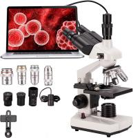
What Can A Light Microscope See ?
A light microscope can see a wide range of objects and structures, including cells, tissues, and microorganisms. It allows for the visualization of various cellular components such as cell membranes, nuclei, and organelles. Additionally, it can be used to observe the morphology and behavior of bacteria, fungi, algae, and other small organisms. Light microscopes are commonly used in biological research, medicine, and education to study the structure and function of living organisms at the cellular level. However, it is important to note that the resolution of a light microscope is limited, typically around 200 nanometers, which means it cannot visualize smaller structures such as individual molecules or viruses.
1、 Cellular structures and organelles
A light microscope, also known as an optical microscope, is a widely used tool in biology and other scientific fields. It uses visible light and lenses to magnify and observe specimens. While it has limitations in terms of resolution and magnification compared to more advanced techniques like electron microscopy, a light microscope can still provide valuable insights into cellular structures and organelles.
With a light microscope, one can observe various cellular structures such as the cell membrane, nucleus, cytoplasm, and cell wall (in plant cells). It allows scientists to study the general morphology and organization of cells, as well as the arrangement and movement of organelles within them. For example, one can observe the shape and size of the nucleus, the presence of membrane-bound organelles like mitochondria and endoplasmic reticulum, and the network of microtubules and microfilaments that make up the cytoskeleton.
In recent years, advancements in light microscopy techniques have further expanded the capabilities of light microscopes. For instance, the development of fluorescence microscopy has enabled the visualization of specific molecules and structures within cells. By labeling cellular components with fluorescent markers, scientists can track the movement of proteins, observe cellular processes in real-time, and study the interactions between different organelles.
Additionally, techniques like confocal microscopy and super-resolution microscopy have improved the resolution of light microscopes, allowing for the visualization of smaller structures and details within cells. These advancements have revolutionized the field of cell biology, enabling researchers to delve deeper into the intricacies of cellular structures and organelles.
In conclusion, a light microscope can see cellular structures and organelles, providing valuable information about the morphology and organization of cells. With recent advancements in light microscopy techniques, scientists can now study cellular processes and interactions at a more detailed level, enhancing our understanding of the complex world within cells.
2、 Tissue organization and architecture
A light microscope is a powerful tool used in biology and medicine to visualize and study various biological specimens. It can provide valuable insights into the tissue organization and architecture of living organisms. With its ability to magnify objects up to 1000 times, a light microscope allows scientists to observe and analyze the intricate details of tissues.
Using a light microscope, researchers can examine the arrangement and structure of cells within tissues. They can observe the different types of cells present, their organization, and how they interact with each other. This information is crucial for understanding the function and behavior of tissues in both healthy and diseased states.
Furthermore, a light microscope can reveal the overall architecture of tissues, including the arrangement of blood vessels, connective tissue, and other structural components. This knowledge is essential for studying the development, growth, and regeneration of tissues, as well as for diagnosing and treating various diseases.
In recent years, advancements in light microscopy techniques have further enhanced our understanding of tissue organization and architecture. For example, confocal microscopy allows for the visualization of tissues in three dimensions, providing a more comprehensive view of their structure. Additionally, the development of fluorescent labeling techniques enables the specific visualization of certain molecules or cell types within tissues, allowing for more detailed analysis.
In conclusion, a light microscope can see and reveal the tissue organization and architecture of living organisms. It provides valuable information about cell arrangement, cell types, and overall tissue structure. With the latest advancements in microscopy techniques, scientists can now obtain even more detailed and comprehensive insights into the complex world of tissues.
3、 Microorganisms and bacteria
A light microscope is a powerful tool that allows scientists to observe and study various biological specimens. It uses visible light to magnify and resolve the details of the sample being observed. While the resolution of a light microscope is limited compared to electron microscopes, it can still provide valuable insights into the world of microorganisms and bacteria.
Microorganisms, such as bacteria, fungi, and protozoa, can be observed using a light microscope. These organisms are typically too small to be seen with the naked eye, but the magnification capabilities of a light microscope allow scientists to study their structure, behavior, and interactions. For example, bacteria can be observed to determine their shape, size, and arrangement, which can provide important information about their classification and potential pathogenicity.
In recent years, advancements in light microscopy techniques have further expanded the capabilities of this tool. Fluorescence microscopy, for instance, allows scientists to label specific molecules or structures within microorganisms and bacteria with fluorescent dyes. This enables the visualization of specific cellular components, such as DNA, proteins, or organelles, providing a deeper understanding of their functions and interactions.
Additionally, techniques like phase contrast microscopy and differential interference contrast microscopy enhance the contrast and visibility of transparent or unstained samples, making it easier to observe delicate structures or live microorganisms in their natural state.
In conclusion, a light microscope can see microorganisms and bacteria, providing valuable information about their structure, behavior, and interactions. With the latest advancements in microscopy techniques, scientists can now delve even deeper into the microscopic world, unraveling the mysteries of these tiny organisms and their role in various biological processes.
4、 Blood cells and other bodily components
A light microscope is a powerful tool used in various scientific fields to observe and study microscopic objects. It uses visible light to magnify and resolve small structures that are otherwise invisible to the naked eye. When it comes to what a light microscope can see, one of the most notable applications is the examination of blood cells and other bodily components.
Blood cells, including red blood cells, white blood cells, and platelets, can be easily observed and analyzed using a light microscope. This allows scientists and medical professionals to study their morphology, count their numbers, and identify any abnormalities or diseases. Additionally, a light microscope can also reveal other bodily components such as epithelial cells, muscle fibers, and connective tissues.
However, it is important to note that the capabilities of a light microscope have certain limitations. Due to the diffraction of light, the resolution of a light microscope is limited to around 200 nanometers. This means that structures smaller than this limit may not be clearly visible or distinguishable. For example, viruses, bacteria, and subcellular organelles like mitochondria or ribosomes are generally beyond the resolution of a light microscope.
To overcome these limitations, scientists have developed more advanced microscopy techniques such as electron microscopy, confocal microscopy, and super-resolution microscopy. These techniques allow for higher magnification and resolution, enabling the visualization of smaller structures and providing more detailed insights into cellular and molecular processes.
In conclusion, a light microscope is a valuable tool for observing and studying blood cells and other bodily components. While it has its limitations in terms of resolution, it remains an essential instrument in many scientific and medical research settings.
