What Can I See With A 1000x Microscope ?
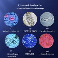
With a 1000x microscope, you can see a wide range of microscopic objects and details. You can observe cells, bacteria, fungi, and other microorganisms in great detail. Additionally, you can examine the structure of plant and animal tissues, such as the arrangement of cells in leaves or the organization of muscle fibers. You may also be able to visualize the intricate patterns and textures of minerals and crystals. Furthermore, you can explore the world of microorganisms in pond water, discovering various protozoa, algae, and other tiny creatures. Overall, a 1000x microscope allows you to delve into the fascinating realm of the microscopic world and explore its diverse wonders.
1、 Cellular Structures and Organelles
With a 1000x microscope, you can observe a wide range of cellular structures and organelles in great detail. This level of magnification allows for the visualization of intricate features within cells that are otherwise invisible to the naked eye.
One of the most prominent structures you can observe is the cell nucleus. The nucleus contains the cell's genetic material, DNA, and is surrounded by a nuclear membrane. Within the nucleus, you can see the nucleolus, which is responsible for the production of ribosomes.
Another important organelle that becomes visible at this magnification is the mitochondrion. These organelles are often referred to as the "powerhouses" of the cell as they generate energy through cellular respiration. With a 1000x microscope, you can observe the inner membrane folds of mitochondria, known as cristae, which increase the surface area for energy production.
Additionally, you can observe other organelles such as the endoplasmic reticulum (ER), Golgi apparatus, and lysosomes. The ER is involved in protein synthesis and lipid metabolism, and its structure can be seen as a network of interconnected tubules and sacs. The Golgi apparatus is responsible for modifying, sorting, and packaging proteins for transport within the cell or secretion outside of the cell. Lysosomes, on the other hand, contain enzymes that break down waste materials and cellular debris.
Furthermore, a 1000x microscope allows for the observation of cellular structures like the cytoskeleton, which provides structural support and facilitates cell movement. You can see microtubules, microfilaments, and intermediate filaments that make up the cytoskeleton.
It is important to note that the latest advancements in microscopy techniques, such as super-resolution microscopy, have pushed the boundaries of what can be observed at high magnifications. These techniques have allowed scientists to visualize cellular structures and organelles with even greater detail, revealing previously unknown substructures and molecular interactions.

2、 Microorganisms and Bacteria
With a 1000x microscope, you can observe a wide range of microorganisms and bacteria that are otherwise invisible to the naked eye. This level of magnification allows for detailed examination of their structures, behaviors, and interactions. Microorganisms are diverse and abundant, and they play crucial roles in various ecosystems, including our own bodies.
One of the most commonly observed microorganisms are bacteria. Bacteria are single-celled organisms that exist in countless shapes, sizes, and arrangements. They can be found virtually everywhere, from soil and water to the human gut. Under a 1000x microscope, you can see the intricate details of bacterial cells, such as their cell walls, flagella (used for movement), and pili (used for attachment). You can also observe their reproductive structures, such as spores or budding.
Beyond bacteria, a 1000x microscope allows you to explore other microorganisms like protozoa, algae, and fungi. Protozoa are single-celled eukaryotes that exhibit remarkable diversity in their shapes and locomotion methods. Algae, on the other hand, are photosynthetic microorganisms that can range from unicellular to multicellular forms. Fungi, including molds and yeasts, can also be observed at this magnification, revealing their filamentous structures and reproductive spores.
It is important to note that the field of microbiology is constantly evolving, and new discoveries are being made regularly. For instance, recent advancements in microscopy techniques, such as super-resolution microscopy, have allowed scientists to observe even smaller structures within microorganisms. These advancements have provided insights into the intricate workings of microbial cells, including their internal organelles and molecular processes.
In conclusion, a 1000x microscope offers a fascinating glimpse into the world of microorganisms and bacteria. It allows us to appreciate the complexity and diversity of these tiny life forms and provides valuable insights into their structures and behaviors. As technology continues to advance, our understanding of microorganisms and their significance in various fields, such as medicine and ecology, will undoubtedly expand.

3、 Tissue and Cell Morphology
With a 1000x microscope, you can observe and study various aspects of tissue and cell morphology in great detail. This level of magnification allows for a closer examination of cellular structures and their organization within tissues.
At this magnification, you can observe the intricate details of cell membranes, cytoplasmic components, and organelles such as mitochondria, Golgi apparatus, and endoplasmic reticulum. You can also study the nucleus, including the chromatin structure and nucleoli. Additionally, you can observe the arrangement and interactions of different cell types within tissues, providing insights into their organization and function.
Furthermore, a 1000x microscope enables the observation of cellular processes such as mitosis and meiosis, allowing for the study of cell division and genetic material distribution. This level of magnification is also useful for examining cellular abnormalities, such as cancerous cells or cellular damage caused by diseases or external factors.
It is important to note that the latest advancements in microscopy techniques, such as confocal microscopy and super-resolution microscopy, have further enhanced our understanding of tissue and cell morphology. These techniques provide even higher resolution and three-dimensional imaging capabilities, allowing for the visualization of subcellular structures and dynamic processes within living cells.
In conclusion, a 1000x microscope provides a powerful tool for studying tissue and cell morphology. It allows for detailed examination of cellular structures, organization within tissues, cellular processes, and abnormalities. However, it is worth exploring the latest microscopy techniques to further expand our understanding of the complex world of cells and tissues.

4、 Blood Cells and Circulatory System
With a 1000x microscope, you can observe a wide range of biological structures and processes, including the intricate world of blood cells and the circulatory system. This level of magnification allows for detailed examination of these components, providing valuable insights into their structure and function.
When observing blood cells, you can see the three main types: red blood cells (erythrocytes), white blood cells (leukocytes), and platelets (thrombocytes). Red blood cells appear as small, biconcave discs, responsible for carrying oxygen throughout the body. White blood cells, on the other hand, come in various shapes and sizes, and play a crucial role in the immune response, defending the body against pathogens. Platelets are tiny cell fragments involved in blood clotting.
Furthermore, a 1000x microscope enables you to explore the circulatory system in more detail. You can observe the structure of blood vessels, including arteries, veins, and capillaries. Arteries carry oxygenated blood away from the heart, while veins transport deoxygenated blood back to the heart. Capillaries, the smallest blood vessels, facilitate the exchange of oxygen, nutrients, and waste products between the blood and surrounding tissues.
In recent years, advancements in microscopy techniques have allowed for even more detailed observations. For instance, with the use of fluorescent dyes and confocal microscopy, it is possible to visualize specific components within blood cells, such as hemoglobin in red blood cells or different types of granules in white blood cells. Additionally, techniques like intravital microscopy enable the study of blood flow dynamics and interactions between blood cells and the vessel wall in real-time.
In conclusion, a 1000x microscope provides a fascinating view of blood cells and the circulatory system. It allows for the observation of their structure, function, and interactions, contributing to our understanding of various physiological processes and potential abnormalities.


