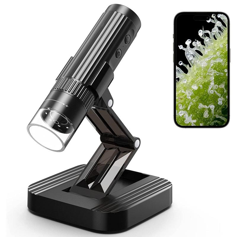
What Can You Look At Under A Microscope ?
Under a microscope, you can look at a wide range of specimens, including cells, tissues, microorganisms, crystals, and other small objects. With a light microscope, you can observe living or fixed specimens, such as bacteria, fungi, algae, and protozoa, as well as plant and animal cells and tissues. You can also examine non-living materials, such as minerals, fibers, and polymers, to study their structure and properties.
With an electron microscope, you can achieve much higher magnification and resolution, allowing you to see even smaller structures, such as viruses, molecules, and atoms. You can also use specialized techniques, such as scanning electron microscopy and transmission electron microscopy, to study the surface and internal structure of materials at high resolution.
Overall, microscopy is a powerful tool for scientific research, allowing us to explore the microscopic world and gain insights into the structure and function of living and non-living systems.
1、 Cells and Tissues
What can you look at under a microscope? Cells and tissues are some of the most common specimens that can be observed under a microscope. Cells are the basic building blocks of life, and they can be found in all living organisms. Tissues, on the other hand, are groups of cells that work together to perform a specific function.
Under a microscope, cells can be observed in great detail, allowing scientists to study their structure, function, and behavior. This has led to many important discoveries in the fields of biology, medicine, and genetics. For example, the discovery of the structure of DNA was made possible by observing cells under a microscope.
Tissues can also be observed under a microscope, allowing scientists to study their structure and function. This has led to many important discoveries in the fields of medicine and biology. For example, the study of cancer tissues has led to the development of new treatments for cancer.
In recent years, advances in microscopy technology have allowed scientists to observe cells and tissues in even greater detail. For example, confocal microscopy allows scientists to observe cells in three dimensions, while super-resolution microscopy allows them to observe structures that were previously too small to be seen.
In conclusion, cells and tissues are some of the most important specimens that can be observed under a microscope. They have led to many important discoveries in the fields of biology, medicine, and genetics, and advances in microscopy technology continue to expand our understanding of these fundamental building blocks of life.

2、 Microorganisms
What can you look at under a microscope? One of the most fascinating things to observe under a microscope is microorganisms. These tiny living organisms are too small to be seen with the naked eye, but under a microscope, they reveal a whole new world of complexity and diversity.
Microorganisms include bacteria, viruses, fungi, and protozoa. They are found in almost every environment on Earth, from soil and water to the human body. Some microorganisms are beneficial, such as those that help us digest food or produce antibiotics. Others can be harmful, causing diseases like pneumonia, tuberculosis, and COVID-19.
Recent advances in microscopy technology have allowed scientists to study microorganisms in greater detail than ever before. For example, electron microscopy can reveal the ultrastructure of cells and viruses, while fluorescence microscopy can track the movement of individual molecules within cells.
One of the most exciting areas of research in microbiology is the study of the human microbiome. This refers to the collection of microorganisms that live on and inside our bodies, which play a crucial role in our health and wellbeing. By studying the microbiome, scientists hope to develop new treatments for diseases like obesity, diabetes, and inflammatory bowel disease.
In conclusion, microorganisms are a fascinating subject to study under a microscope. They are incredibly diverse and complex, and recent advances in microscopy technology have allowed us to study them in greater detail than ever before. As our understanding of microorganisms continues to grow, we may be able to unlock new treatments and cures for a wide range of diseases.

3、 Blood and Body Fluids
What can you look at under a microscope? Blood and body fluids are some of the most commonly examined specimens under a microscope. Blood is a complex fluid that contains various types of cells, including red blood cells, white blood cells, and platelets. These cells can be examined under a microscope to identify any abnormalities or diseases. For example, red blood cells can be examined to determine their size, shape, and color, which can help diagnose conditions such as anemia or sickle cell disease. White blood cells can be examined to identify infections or immune system disorders.
Body fluids such as urine, cerebrospinal fluid, and synovial fluid can also be examined under a microscope. Urine can be examined to identify the presence of bacteria, blood, or other substances that may indicate a urinary tract infection or kidney disease. Cerebrospinal fluid can be examined to diagnose conditions such as meningitis or multiple sclerosis. Synovial fluid can be examined to diagnose joint diseases such as rheumatoid arthritis.
In recent years, advances in microscopy technology have allowed for even more detailed examination of blood and body fluids. For example, high-resolution microscopy techniques such as confocal microscopy and super-resolution microscopy can provide detailed images of cells and structures within cells. These techniques have been used to study the structure and function of blood cells and to develop new diagnostic tools for diseases such as cancer.
In conclusion, blood and body fluids are important specimens that can be examined under a microscope to diagnose a wide range of diseases and conditions. Advances in microscopy technology continue to improve our understanding of these specimens and their role in health and disease.

4、 Minerals and Crystals
What can you look at under a microscope? Minerals and crystals are some of the most fascinating objects to observe under a microscope. With the advancement of technology, scientists can now study the atomic structure of minerals and crystals in great detail, providing insights into their physical and chemical properties.
Under a microscope, minerals and crystals can reveal a range of features, including their color, shape, and texture. By examining the crystal lattice structure, scientists can determine the mineral's chemical composition and its properties, such as hardness, cleavage, and fracture.
Recent developments in microscopy have allowed scientists to study minerals and crystals at the nanoscale level. This has led to the discovery of new minerals and the development of new materials with unique properties. For example, researchers have used microscopy to study the structure of zeolites, a type of mineral with a porous structure that can be used for catalysis and gas separation.
In addition to scientific research, microscopy has also been used in the field of gemology to identify and authenticate gemstones. By examining the internal structure of a gemstone, gemologists can determine its authenticity and origin.
Overall, minerals and crystals are fascinating objects to observe under a microscope, providing insights into their physical and chemical properties and leading to new discoveries in materials science and other fields.


