What Can You See Through A Light Microscope ?
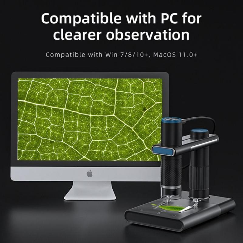
A light microscope is an optical instrument that uses visible light and lenses to magnify small objects. It can be used to see a variety of specimens, including cells, tissues, bacteria, and small organisms. With a light microscope, you can see the internal structures of cells, such as the nucleus, mitochondria, and cytoplasm. You can also observe the shape and size of cells, as well as their arrangement in tissues. Bacteria can be seen as individual cells or in colonies, and their shape and arrangement can provide clues to their identity. Small organisms, such as protozoa and algae, can also be observed with a light microscope. Overall, a light microscope is a valuable tool for studying the microscopic world and understanding the structure and function of living organisms.
1、 Cellular structures

A light microscope is a powerful tool that allows scientists to observe cellular structures. With this microscope, scientists can see the basic structures of cells, such as the cell membrane, nucleus, and cytoplasm. They can also observe the organelles within the cell, such as mitochondria, ribosomes, and the endoplasmic reticulum.
In recent years, advances in light microscopy have allowed scientists to see even more detailed cellular structures. For example, they can now observe the movement of molecules within cells, as well as the interactions between different cellular structures. They can also observe the structure of proteins and other molecules within cells, which can provide important insights into how cells function.
One of the most exciting developments in light microscopy is the ability to observe living cells in real-time. This has allowed scientists to study cellular processes as they happen, such as cell division, protein synthesis, and the movement of molecules within cells. This has led to a better understanding of how cells function and has opened up new avenues for research into diseases such as cancer and Alzheimer's.
Overall, the light microscope is an essential tool for studying cellular structures. With advances in technology, scientists are able to see more and more detailed structures within cells, which is leading to a better understanding of how cells function and how they can be targeted for therapeutic purposes.
2、 Microorganisms

What can you see through a light microscope? One of the most common things that can be seen through a light microscope are microorganisms. These are tiny living organisms that are too small to be seen with the naked eye. Examples of microorganisms include bacteria, viruses, fungi, and protozoa.
When viewed under a light microscope, microorganisms can reveal a wealth of information about their structure, behavior, and function. For example, bacteria can be observed in their various shapes and sizes, and their cell walls and internal structures can be studied. Viruses can be seen as tiny particles, and their shapes and structures can be analyzed. Fungi can be observed in their various forms, including spores, hyphae, and fruiting bodies. Protozoa can be seen swimming and moving around, and their internal structures can be studied.
In recent years, advances in microscopy technology have allowed scientists to see microorganisms in even greater detail. For example, confocal microscopy and electron microscopy have enabled researchers to study microorganisms at the cellular and molecular level, revealing new insights into their biology and behavior.
Overall, the study of microorganisms through light microscopy has been instrumental in advancing our understanding of the microbial world and its impact on human health, the environment, and other areas of science.
3、 Tissues and organs

What can you see through a light microscope? Tissues and organs are some of the structures that can be observed using this type of microscope. Light microscopes use visible light to magnify and illuminate specimens, allowing scientists to study their structure and function.
When examining tissues, light microscopes can reveal the arrangement of cells and their various components, such as nuclei, cytoplasm, and organelles. This information can be used to identify different types of tissues, such as muscle, nerve, or epithelial tissue. Light microscopes can also be used to study the structure of organs, such as the liver, kidney, or heart. By examining the arrangement of cells and tissues within an organ, scientists can gain insight into its function and how it interacts with other organs in the body.
Recent advances in light microscopy have expanded the capabilities of this technology. For example, confocal microscopy uses lasers to create high-resolution images of specimens, allowing scientists to study the three-dimensional structure of tissues and organs in greater detail. Fluorescence microscopy uses fluorescent dyes to label specific structures within cells, making them easier to visualize and study. These techniques have revolutionized our understanding of biological structures and processes, and continue to be important tools in modern research.
In conclusion, light microscopes are powerful tools for studying tissues and organs. They allow scientists to observe the structure and function of these structures, and recent advances in microscopy have expanded their capabilities even further. By using light microscopes to study tissues and organs, scientists can gain insight into the complex workings of the human body and develop new treatments for diseases and disorders.
4、 Blood cells

Through a light microscope, one can see a variety of biological specimens, including blood cells. Blood cells are the cellular components of blood, which are responsible for carrying oxygen, fighting infections, and regulating the body's immune response. There are three types of blood cells: red blood cells, white blood cells, and platelets.
Red blood cells, also known as erythrocytes, are the most abundant type of blood cell. They are responsible for carrying oxygen from the lungs to the body's tissues and removing carbon dioxide from the body. Under a light microscope, red blood cells appear as small, biconcave discs with a diameter of approximately 7 micrometers.
White blood cells, also known as leukocytes, are responsible for fighting infections and foreign invaders in the body. There are several types of white blood cells, including neutrophils, lymphocytes, monocytes, eosinophils, and basophils. Under a light microscope, white blood cells appear as irregularly shaped cells with a diameter of approximately 10-20 micrometers.
Platelets, also known as thrombocytes, are responsible for blood clotting. Under a light microscope, platelets appear as small, irregularly shaped cells with a diameter of approximately 2-4 micrometers.
It is important to note that while a light microscope can provide valuable information about blood cells, it has its limitations. For example, it cannot provide information about the internal structures of cells or their molecular composition. Therefore, other techniques such as electron microscopy and molecular biology techniques are often used in conjunction with light microscopy to gain a more complete understanding of blood cells and their functions.


