What Can You See With 400x Microscope ?
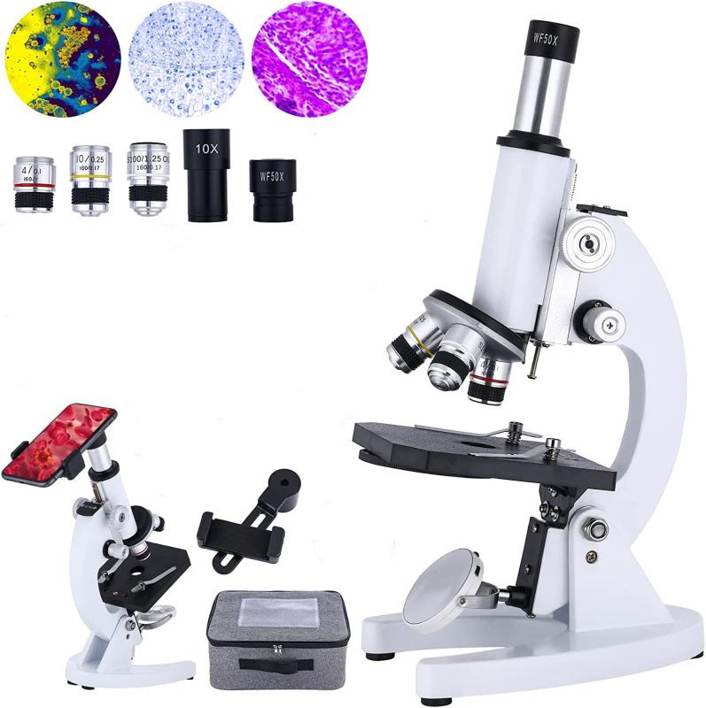
With a 400x microscope, you can see a wide range of microscopic objects and details. This level of magnification allows you to observe cells, bacteria, fungi, and other microorganisms in greater detail. You can also examine the structure of plant and animal tissues, including individual cells and their components. Additionally, you can observe the fine details of small organisms such as protozoa and algae. With a 400x microscope, you can explore the intricate world of microscopic life and gain a deeper understanding of their structures and functions.
1、 Cellular structures and organelles in greater detail
With a 400x microscope, you can observe cellular structures and organelles in greater detail. This level of magnification allows for a closer examination of the intricate components that make up a cell. For example, you can observe the nucleus, mitochondria, endoplasmic reticulum, Golgi apparatus, and various other organelles within a cell.
The nucleus, often referred to as the control center of the cell, contains the genetic material and can be observed with greater clarity at 400x magnification. The mitochondria, responsible for energy production, can also be studied in more detail, allowing for a better understanding of their structure and function.
Furthermore, the endoplasmic reticulum, involved in protein synthesis and lipid metabolism, can be observed with increased resolution. The Golgi apparatus, responsible for modifying, sorting, and packaging proteins, can also be examined in greater detail, providing insights into its complex structure.
It is important to note that advancements in microscopy techniques and technologies have allowed for even higher magnifications and resolutions. For instance, with the advent of super-resolution microscopy, scientists can now visualize cellular structures and organelles at the nanoscale level, providing unprecedented details about their organization and interactions.
In conclusion, a 400x microscope enables the observation of cellular structures and organelles in greater detail, enhancing our understanding of their morphology and function. However, it is worth noting that the field of microscopy continues to evolve, and newer techniques offer even more precise and detailed views of cellular components.

2、 Microorganisms such as bacteria and protozoa
With a 400x microscope, you can observe a wide range of microorganisms such as bacteria and protozoa. These microorganisms are too small to be seen with the naked eye, but with the magnification power of a 400x microscope, they become visible and their structures can be studied in detail.
Bacteria are single-celled organisms that exist in various shapes and sizes. With a 400x microscope, you can observe the different morphologies of bacteria, including rod-shaped (bacilli), spherical (cocci), and spiral-shaped (spirilla). You can also observe the presence of flagella, which are whip-like structures that some bacteria use for movement. Additionally, you can study the arrangement of bacteria in colonies or clusters, which can provide insights into their growth patterns and behavior.
Protozoa, on the other hand, are eukaryotic microorganisms that are larger and more complex than bacteria. With a 400x microscope, you can observe the various structures within protozoa, such as the nucleus, cytoplasm, and specialized organelles like contractile vacuoles or cilia. You can also observe their movement patterns, as many protozoa have unique modes of locomotion, such as flagella, cilia, or pseudopodia.
It is important to note that the field of microbiology is constantly evolving, and new discoveries are being made. With advancements in microscopy techniques and technology, scientists are now able to observe microorganisms at even higher magnifications and resolutions. This allows for a more detailed understanding of their structures, interactions, and functions. Therefore, while a 400x microscope can provide valuable insights into the world of microorganisms, it is just one tool in the vast arsenal of microbiologists.

3、 Tissue samples and their cellular composition
With a 400x microscope, one can observe tissue samples and their cellular composition in great detail. This level of magnification allows for the examination of various types of tissues, such as epithelial, connective, muscular, and nervous tissues. By studying these tissues, scientists and researchers can gain insights into their structure, organization, and function.
At 400x magnification, one can observe the individual cells that make up the tissue. This includes the cell membrane, nucleus, cytoplasm, and any specialized structures or organelles within the cell. By examining the cellular composition, scientists can identify different cell types and their specific roles within the tissue. For example, in epithelial tissue, one can observe the tightly packed cells that form a protective barrier, while in muscular tissue, one can observe the elongated muscle fibers responsible for contraction.
Furthermore, a 400x microscope can reveal important details about the arrangement and organization of cells within the tissue. This can provide insights into how cells interact and communicate with each other, as well as how they contribute to the overall function of the tissue. For instance, in nervous tissue, one can observe the branching extensions of neurons and their connections, which are crucial for transmitting electrical signals.
It is important to note that the latest advancements in microscopy techniques, such as confocal microscopy and super-resolution microscopy, have further enhanced our ability to study tissue samples. These techniques allow for even higher magnification and resolution, enabling researchers to visualize subcellular structures and processes with greater clarity.
In conclusion, a 400x microscope is a valuable tool for studying tissue samples and their cellular composition. It allows for the observation of individual cells, their structures, and their organization within the tissue. However, it is worth noting that the latest microscopy techniques have pushed the boundaries of what can be seen and understood at the cellular level, providing even more detailed insights into tissue biology.

4、 Fine details of plant structures like stomata and trichomes
With a 400x microscope, you can observe and study the fine details of plant structures such as stomata and trichomes. Stomata are tiny openings found on the surface of leaves and stems that allow for gas exchange, including the intake of carbon dioxide and the release of oxygen. Trichomes, on the other hand, are hair-like structures that can be found on the surface of plants, serving various functions such as protection against herbivores, reducing water loss, and reflecting excess sunlight.
At 400x magnification, you can clearly observe the intricate features of stomata and trichomes. Stomata appear as small, oval-shaped openings surrounded by specialized cells known as guard cells. By examining stomata under a microscope, scientists can analyze their density, size, and arrangement, which can provide insights into a plant's adaptation to different environmental conditions.
Trichomes, which come in various shapes and sizes, can also be examined in detail with a 400x microscope. These structures can be unicellular or multicellular and may have different functions depending on the plant species. By studying trichomes, researchers can gain a better understanding of their role in plant defense mechanisms, as well as their potential applications in fields such as agriculture and medicine.
It is important to note that the capabilities of microscopes have significantly advanced in recent years. With the latest advancements in microscopy technology, such as high-resolution imaging and digital microscopy, scientists can now capture even finer details of plant structures at higher magnifications. These advancements allow for more precise analysis and contribute to a deeper understanding of the intricate world of plants.


There are no comments for this blog.