What Does A Microscope Look Like ?
A microscope is a scientific instrument used to magnify and observe small objects or organisms that are not visible to the naked eye. It typically consists of a base, an adjustable stage, and a tube with lenses. The base provides stability and support for the microscope. The adjustable stage is where the specimen is placed for observation and can be moved in different directions to position the specimen under the lenses. The tube contains the optical system, which includes the eyepiece and objective lenses. The eyepiece is where the viewer looks through to observe the magnified image, while the objective lenses are responsible for magnifying the specimen. Microscopes can have different designs and variations, such as compound microscopes with multiple lenses or stereo microscopes with two eyepieces for a three-dimensional view.
1、 Optical Microscope: Traditional microscope using lenses to magnify specimens.
A microscope is a scientific instrument that is used to magnify small objects or specimens that are not visible to the naked eye. The most common type of microscope is the optical microscope, which uses lenses to magnify the specimen.
An optical microscope typically consists of several key components. The most prominent part is the body or frame, which holds all the other parts together. Attached to the body is the stage, where the specimen is placed for observation. The stage usually has clips or holders to secure the specimen in place.
Above the stage, there is a light source that illuminates the specimen. In traditional microscopes, this light source is usually a bulb or a mirror that reflects light onto the specimen. However, with advancements in technology, LED lights are now commonly used as they provide a more consistent and brighter illumination.
The lenses are the most important part of the microscope. There are usually two sets of lenses - the objective lenses and the eyepiece or ocular lens. The objective lenses are located on a rotating turret and can be switched to provide different levels of magnification. The eyepiece lens is located at the top of the microscope and is where the viewer looks through to observe the magnified specimen.
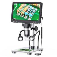
In recent years, there have been significant advancements in microscope technology. Digital microscopes have become increasingly popular, allowing for the capture and analysis of images on a computer screen. Additionally, there are now portable and handheld microscopes that are compact and easy to use in the field.
Overall, while the basic design of an optical microscope has remained relatively unchanged, advancements in technology have allowed for improved functionality, ease of use, and more precise observations.

2、 Electron Microscope: Uses electron beams for high-resolution imaging.
A microscope is a scientific instrument that is used to magnify and observe objects that are too small to be seen with the naked eye. It allows scientists and researchers to study the intricate details of various specimens, such as cells, bacteria, and other microorganisms.
There are different types of microscopes, each with its own unique features and capabilities. One of the most advanced and powerful types is the electron microscope. Unlike traditional light microscopes that use visible light to illuminate the specimen, electron microscopes use a beam of electrons for imaging. This allows for much higher resolution and magnification, enabling scientists to see even smaller details.
An electron microscope consists of several main components. It includes an electron gun that produces a beam of electrons, electromagnetic lenses that focus and control the electron beam, a specimen chamber where the specimen is placed, and a detector that captures the electrons that pass through or scatter from the specimen. The microscope also has a computer system for image processing and analysis.
In recent years, there have been advancements in electron microscopy technology. For example, the development of aberration-corrected electron microscopy has significantly improved the resolution and clarity of images. Additionally, the integration of scanning electron microscopy (SEM) and transmission electron microscopy (TEM) techniques has allowed for a more comprehensive understanding of the structure and composition of specimens.
Overall, electron microscopes have revolutionized the field of microscopy by providing scientists with a powerful tool to explore the microscopic world in unprecedented detail. They have contributed to numerous scientific discoveries and continue to be an essential instrument in various fields, including biology, materials science, and nanotechnology.

3、 Confocal Microscope: Provides detailed 3D images using laser scanning.
A confocal microscope is a powerful tool used in scientific research and medical diagnostics. It is designed to provide detailed 3D images of biological samples using laser scanning technology. The microscope consists of several key components that work together to produce high-resolution images.
The main part of a confocal microscope is the laser scanning system. It uses a laser beam to illuminate the sample, and the light emitted from the sample is then collected by a detector. The laser scanning system allows for precise control over the focal plane, enabling the capture of images at different depths within the sample. This feature is particularly useful for studying thick samples or tissues.
Another important component of a confocal microscope is the pinhole aperture. This aperture is placed in front of the detector and allows only the light emitted from the focal plane to pass through. By blocking out-of-focus light, the pinhole aperture enhances the image contrast and improves the overall resolution of the microscope.
In recent years, there have been advancements in confocal microscopy technology. For example, the development of super-resolution techniques has allowed researchers to achieve even higher resolution images. These techniques, such as stimulated emission depletion (STED) microscopy and structured illumination microscopy (SIM), have pushed the boundaries of what can be observed with a confocal microscope.
Overall, a confocal microscope is a sophisticated instrument that provides researchers with the ability to visualize biological samples in great detail. Its laser scanning system and pinhole aperture work together to produce high-resolution 3D images, making it an invaluable tool in various scientific fields.

4、 Scanning Probe Microscope: Measures surface properties at atomic scale.
A Scanning Probe Microscope (SPM) is a powerful tool used in nanotechnology and materials science to measure surface properties at the atomic scale. Unlike traditional optical microscopes, which use light to magnify objects, an SPM uses a sharp probe to scan the surface of a sample and create a detailed image.
The main component of an SPM is the probe, which is typically a sharp tip made of a conductive material such as silicon or tungsten. This tip is attached to a cantilever, a flexible arm that can move up and down in response to changes in the surface topography. As the probe scans the sample, it interacts with the surface forces, such as van der Waals forces or electrostatic forces, and these interactions are measured to create an image.
The SPM setup also includes a feedback mechanism that maintains a constant distance between the probe and the sample surface. This allows for precise measurements of surface properties, such as roughness, conductivity, or magnetic fields. The feedback mechanism adjusts the position of the probe based on the detected forces, ensuring accurate imaging.
In recent years, advancements in SPM technology have led to the development of various specialized techniques. For example, Atomic Force Microscopy (AFM) is a common SPM technique that measures the forces between the probe and the sample surface to create a high-resolution image. Scanning Tunneling Microscopy (STM) is another technique that measures the flow of electrons between the probe and the sample surface to create atomic-scale images.
Overall, a Scanning Probe Microscope is a sophisticated instrument that allows scientists to explore and understand the properties of materials at the atomic level. Its ability to provide detailed images and measurements has revolutionized the field of nanotechnology and has contributed to numerous scientific discoveries.










There are no comments for this blog.